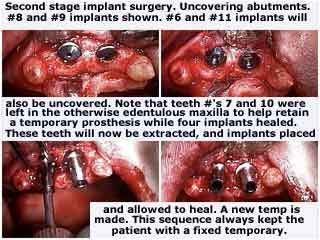
Second stage dental implant surgery. Uncovering the first stage implant abutments. Note: The text in the image says: Second stage implant surgery. Uncovering abutments. #8 and #9 implants shown. #6 and #11 implants will also be uncovered. Note that the teeth #’s 7 and 10 were left in the otherwise edentulous [no teeth] maxilla [upper jaw] to help retain a temporary prosthesis while four implants healed. These teeth will now be extracted, and implants placed and allowed to heal. A new [fixed temporary bridge] is made. This [treatment] sequence always kept the patient with a fixed temporary [bridge].
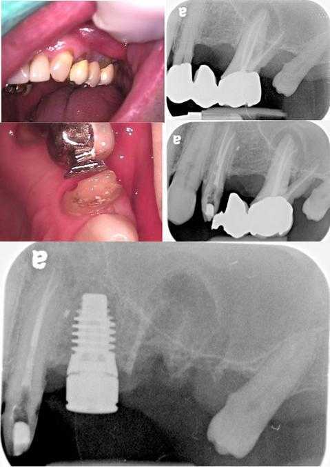
Photos show treatment plan sequencing – all this work was completed in one visit. An old upper left dental bridge #12 – 14 (first row photo & xray) had failed. The crown around #12 was sectioned (second row photo & xray) to facilitate access for the Endodontist. The Oral Surgeon then removed the remaining teeth bridge #13 – 14 and placed a dental implant in the #13 space (third row x-ray). A fixed temporary teeth bridge was then placed from #12 -15 during healing. Treatment sequence logic: 1. Facilitate Endodontist access for root canal, 2. No need to section and remove the bridge from tooth #14 since it would come out with the tooth extraction, 3. Non-bleeding procedures should be performed before those that result in bleeding, 4. Dental implant placement after tooth extraction.
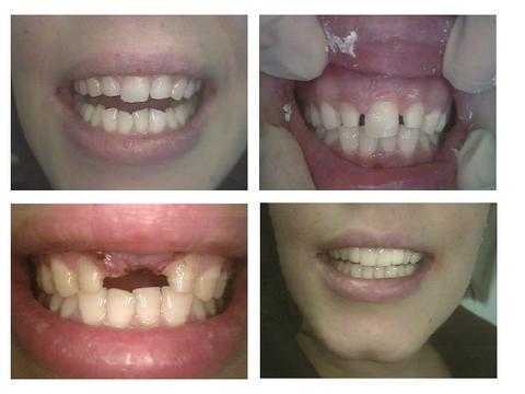
How to plan treatment sequencing – the order of dental treatment. Cosmetic Dentistry for a pretty 27 year old woman from Ireland with only one upper front tooth. How to pictures. The upper lateral incisor teeth had root canal therapy and posts placed. The laterals were then prepared for a dental bridge but more tooth reduction was taken from the mesial. The mesial of both canine teeth were reduced so that the laterals could be “distalized.” The central incisor tooth was extracted and a bone graft was placed. A temporary lab-processed teeth bridge is shown in this photo.
This patient came from Ireland and stayed from Monday to Friday of one week. Treatment was performed in only four hours on one day with the help of an Endodontist, Oral Surgeon, Joseph Tuil Dental Lab and Cosmetic Dentist.
A second lab-processed temporary bridge, reinforced with Ribbond, was placed before the patient’s departure. She will return from Ireland in three months for fabrication and insertion of the final porcelain dental bridge. This second visit will also occur during one week.
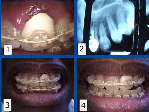
Planning the order of dental treatment involving multi-specialty dentistry. Orthodontic complications – problems with teeth braces. Photos 1) & 2) This patient presented with a failing upper front tooth #9 with an unattractive dental crown. This tooth was maintained for one year by a Periodontist while the Orthodontist straightened her teeth with braces. The #9 tooth root was then extracted from under the crown to allow the gums and bone to heal with about eight months remaining to complete her braces. Pictures 3) & 4) This rootless crown #9 is being held in place only by the orthodontic bracket and wire. This patient preferred to keep this unattractive crown during her braces treatment to minimize simultaneous cosmetic changes, but a cosmetic temporary dental crown could also have been used during this time. A dental implant will be placed about three months following the tooth extraction. The second stage dental implant connection will coincide with the teeth braces removal.
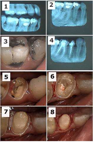
The order or sequence of dental treatment for a patient who presented with tooth pain on her lower right side. The premolar is the focus in this series of photos and x-rays. Root canal, crown build-up and dental crown preparation was performed for both the molar and premolar.
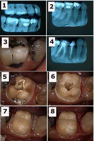
The order or sequence of dental treatment for a patient who presented with tooth pain on her lower right side. The molar is the focus in this series of pictures and xrays. Root canal, crown build-up and tooth crown preparation was performed for both the molar and premolar.
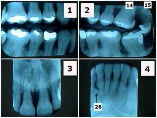
Sequencing dental treatment in a 40-year-old female with severe periodontal gum disease and dental fear anxiety. Discussion with xrays. Note the significant horizontal bone loss and the lack of radiographic x-ray calculus. Tooth #26, adjacent to the vertical bone defect, is probably hopeless. This patient was treated with two rounds of scaling and root planing and then Periodontist reevaluation. Tooth extraction was performed on #15 and #14 had root canal therapy and a tooth crown.
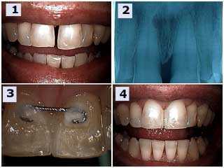
Treatment plan sequence for an upper left front tooth central incisor #9 with significant gum periodontal disease. Before and after. 1) & 2) Pre-operative photo and x-ray radiograph. Note the angular bone defect in the radiograph. The patient reported that the tooth was getting longer and that he never previously had the tooth space gap between his two front teeth. 3) Palatal image of the teeth splint placed between teeth #’s 8 & 9. 4) Post-operative photograph taken one hour later. Teeth bonding placed between the teeth to close the space also hides the palatal splint. The incisal edge of #9 was shortened and the occlusion was checked and adjusted for fremitus. Root planing was next performed and the patient placed on a three-month periodontal reevaluation.
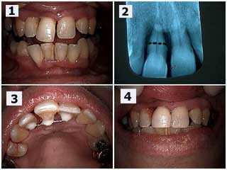
Treatment plan order for a patient who had a very loose upper right central incisor #8. The patient wanted to try to save this tooth as long as possible. Photo 1) The upper incisor tooth has extruded and moved labially (toward the lip) and a diastema – tooth space has occurred. 2) The radiograph shows severe bone loss. The black dotted line shows the location for a potential root resection if necessary. Root planing was performed after a teeth splint was placed. The patient will return in three months to reevaluate periodontal healing with the Periodontist and the potential need for the root resection. Photo 3) Palatal view of the dental splint between teeth #’s 7 – 9. Photo 4) Post-op view on the same day. Notice the diastema teeth space was closed with dental bonding to hide the splint and the incisal edge of #8 was reduced.
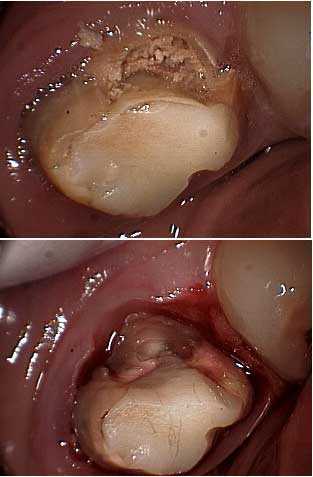
Treatment plan order for tooth decay cavity removal around a molar. Dental caries – tooth decay – has to been removed before referring the patient for crown lengthening periodontal gum surgery so the Periodontist knows how much osseous reduction – bone removal – is necessary.
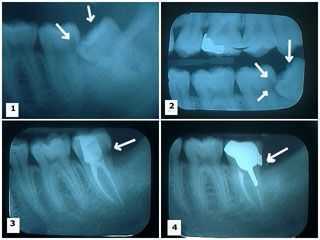
Treatment plan order for wisdom tooth extraction. 1) – 2) Two x-rays show a lower wisdom tooth impacted on an angle – angular bony impaction – and pushing into the adjacent second molar causing a large tooth cavity. 3) This xray shows the wisdom tooth was extracted. Root canal therapy was performed on the remaining second molar with the large distal cavity dental caries still present. 4) A cast gold post and core and dental crown is now in place. Note the distal crown margin completely covers where the distal tooth decay was removed.
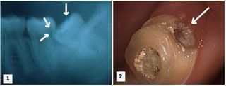
Sequencing dental treatment. 1) This x-ray shows an impacted lower wisdom tooth pushing into the adjacent second molar causing dental caries tooth decay. 2) Tooth decay is present in the distal area of the second molar. Temporary filling material is visible in the occlusal opening following root canal therapy. The wisdom tooth should first be removed before treating the second molar; this is the typical appropriate order of treatment.
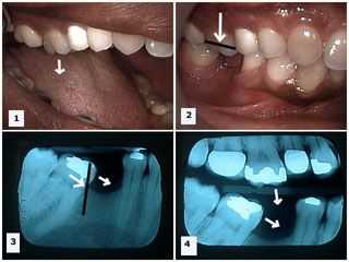
The sequence or order of dental treatment for supraeruption and mesial drift after tooth extraction. Pictures 1) -2) The upper first molar tooth has supraerupted because its opposing antagonist, the lower first molar, has been extracted. X-ray 3) Mesial drift of the lower second molar is seen after removal of the lower first molar. X-ray 4) Both supra-eruption of the upper first molar tooth and mesial drifting of the lower second molar tooth can be seen in this one xray secondary to the removal of the lower first molar. Treatment should first focus on reducing the occlusal height of the supraerupted upper first molar while molar uprighting the lower second molar. Then a dental implant should be placed in the location of the missing tooth. It is not good dentistry to build a dental occlusion around supraeruption.
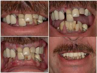
Treatment plan order for oral rehabilitation dental reconstruction. First help this dental phobia anxiety patient overcome their fear. Then create a treatment plan to give them maximum aesthetics and function with minimal discomfort as soon as possible!
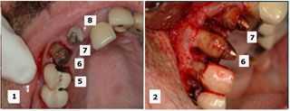
Treatment plan order during selective teeth extraction. The initial treatment plan considered saving teeth #’s 6, 7 and 9 while extracting #’s 5 and 8. During the oral surgery, the Oral Surgeon considered #5 more stable than #6 so #5 was retained and #6 was extracted. The retained teeth will be used to support a fixed temporary teeth bridge until subsequent dental implants are placed.