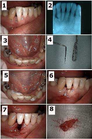
Dental treatment planning a hopeless tooth #26. 1) – 3) Initial photos and x-ray. 4) Photo of the wire used for the teeth splint next to a periodontal probe to give an idea of scale. The wire is doubled to prevent the tooth from rotating around it after placement. 5) Dental bonding the splint to the lingual surfaces of teeth #’s 25 and 27. 6) Initial preparation through the mid-length of the tooth. 7) Following tooth root extraction. 8) The extracted tooth root. This patient has already been through two rounds of scaling and root planing. After healing of this extraction site the Periodontist will perform an apically positioned flap periodontal gum surgery and then possibly a bone and/or soft graft to build up this area. A Maryland bridge with tooth colored wings will be placed after periodontal therapy is completed.
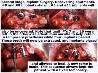
Treatment planning a complete maxillary arch dental implant oral rehabilitation to avoid the use of a removable upper jaw denture. This patient suffered from depression and insisted that she never wear a denture while dental implants healed. These pictures start with the second stage dental implant surgery following osseointegration. Weak teeth #7 and 10 were left in the otherwise edentulous maxilla to help retain a fixed provisional teeth bridge while four upper dental implants osseointegrated. These two teeth will now be extracted and a new provisonal temporary teeth bridge will be made while these remaining dental implants osseointegrate over the next four months. This sequence allows the patient to always have a fixed temporary dental bridge rather than a removable denture while the osseointegration occurs.
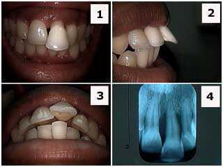
Treatment planning for severe periodontal gum disease in a 24 year old female. 1) Front photo. 2) Side photo. 3) Occlusal picture. 4) Radiograph x-ray. The patient was informed about the severe periodontal condition and that a full mouth Periodontist exam was needed. Teeth braces orthodontics is not appropriate to retract this front tooth because of the severe gum disease. During the case presentation where multiple treatment options were discussed the patient stated her preference to keep these teeth for as long as possible. Treatment option #1 could include scaling and root planning, open flap debridement if necessary, root canal therapy and a dental crown (for aesthetics) and a teeth splint. Treatment option #2 could include just scaling and root planning, incisal adjustment, cosmetic tooth bonding and a teeth splint. Many people prefer to go through an emotional transition with the expected loss of a front tooth.
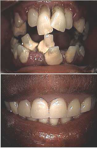
Treatment planning a full mouth oral rehabilitation for a dental phobia fear patient. Before and after pictures – two visits. It is important to produce a dramatic result quickly and comfortably when performing a smile makeover for these patients. Photo #1 of 4.
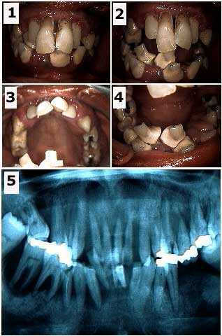
Treatment planning a full mouth dental rehabilitation for a dentist phobia fear patient. Initial visit photos and panoramic x-ray. It is important to determine what teeth, if any, may be saved at least temporarily. It is easier for a patient to emotionally adjust to a temporary prosthesis teeth bridge that has at least some amount of retention provided by natural teeth abutments. The initial abutment teeth chosen were #6, 11, 22 and 28. The decision to fabricate a removable immediate partial denture, rather than a fixed lab-processed provisional teeth bridge, was determined by the particular weakness of tooth #28. The patient was informed that the immediate prosthesis was to be used for the healing phase and that the four remaining teeth abutments, particularly #28, might be subsequently extracted. Photo #2 of 4.
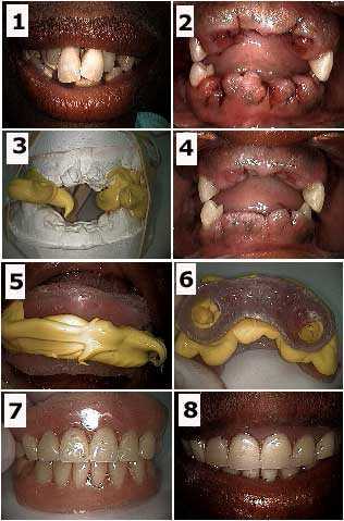
Treatment planning a full mouth smile makeover for a dental phobia anxiety patient. How to pictures. 1) Initial photo. 2) Following teeth extraction of all but the four remaining abutments. 3) Working master models and the teeth bite occlusion made right after the teeth extractions. This will serve for the fabrication of the immediate removable partial dentures. 4) Three days following the extractions. 5) Intra oral photo of the bite rims and bite material. 6) Extra oral photo of bite rims and bite material. The patient did not want any wire clasps to be used to retain the prosthesis. 7) Extra oral photo of the immediate dentures. 8) Intra oral photo of the immediate dentures. A soft reline material was used on the gingival side of these dentures. This soft reline material was allowed to flow into the circular openings (see photo #6) in the acrylic denture as it was seated. This soft reline material provided retention instead of using clasps. This took two visits over three days. Photo #3 of 4.
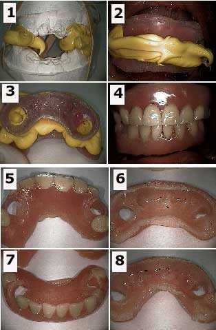
Treatment planning a full mouth oral rehabilitation for a dental anxiety phobia patient. How to pictures 1) Working master models and the teeth bite made right after the teeth extractions. This will serve for the fabrication of the immediate removable partial dentures. 2) Intra oral photo of the bite rims and bite material. 3) Extra oral photo of bite rims and bite material. The patient did not want any wire clasps to be used to retain the prosthesis. 4) Intra-oral view of the immediate dentures. A soft reline material was used on the gingival side of these dentures. This soft reline material was allowed to flow into the circular openings in the acrylic denture as it was seated. This soft reline material provided retention instead of using clasps. 5) & 6) Views of the maxillary denture with soft reline material visible. 7) & 8) Views of the mandibular denture with soft reline material visible. Photo #4 of 4. Developing a treatment plan acceptable to a dental phobia patient is particularly challenging but very rewarding.
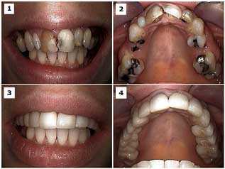
Creating a dental treatment plan for a smile makeover focusing on the upper jaw in a phobic, pretty 33 year-old female. Before and After photos. Treatment included porcelain fused to metal dental crown & bridge, root canal therapy on all teeth abutments, root tip extractions, and facial teeth bonding on both upper lateral incisors. The upper anterior root canal and dental crowns were first completed to show her how pretty her teeth could look and that it could be accomplished quickly and painlessly. Extractions of the hopeless teeth were all completed at one time and initiated early to allow time for wound healing before bridgework. Then root canal therapy was performed on the posterior abutments before their preparation for bridgework. The patient experienced minimal post-operative pain. Treatment plans should be created in a comprehensive and logical order that considers the patients emotions and desire to see results quickly while remaining comfortable. At The Center for Special Dentistry we frequently consult with all appropriate specialists before we start this type of case.
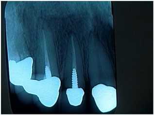
Treatment plan: when not to treat a patient. This lovely woman presented for what she believed would be a quick recementation of a loose upper lateral tooth crown. Many crowns and root canals were recently completed by her local dentist near her home where she was a current patient. There were many reasons why I chose not to treat this patient and instead recommended that she go back to her current dentist for care. 1) It appeared that the crown was completely seated on the post and core instead of being seated on a minimum of two millimeters of sound tooth structure. 2) The post preparation was short and should have been nearly double its length for strength and retention. 3) The post was prefabricated and not cast; this will also decrease its strength and retention. I did not want to take out the loose crown and then find that I also had a loose post and core in my hand. Proper care might indicate a new, longer cast post and core, possible crown lengthening, and a new crown. Crown lengthening surgery could likely affect the crown margins on all the other new anterior crowns. Once you touch it, you own the problem. Both the dentist and patient need to agree what might happen, and what might need to be done, if an “easy” repair turns out to be quite a big problem. This philosophy is both fair and appropriate for the patient and will save a lot of young dentists from having a major, unexpected problem. All things considered it was not worth getting started. The patient was appreciative of my assessment and was happy to return to her dentist for the repair.
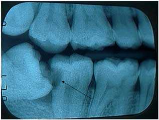
Treatment planning wisdom tooth extraction. This mandibular angular impaction has caused a huge tooth cavity in the second molar that could probably require root canal therapy and a dental crown to repair. The patient did not feel pain in this tooth. A proper treatment plan and case presentation for the extraction of this wisdom tooth should include the work needed on this adjacent tooth.
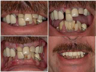
Treatment plan a smile makeover for nervous dentistry patients to get them into a fixed provisional temporary dental bridge quickly and comfortably. This initial treatment took only one visit.
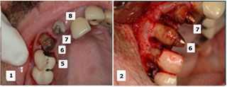
Treatment planning selective teeth extraction during a maxillary dental reconstruction. The initial treatment plan considered saving teeth #’s 6, 7 and 9 while extracting teeth #’s 5 and 8. During the oral surgery the Oral Surgeon decided that tooth #5 was stronger than #6 so #5 was retained and #6 was extracted. The retained teeth will be used to support a fixed temporary dental bridge until subsequent implant dentistry is completed. Write dental treatment plans that allow for changes if appropriate.