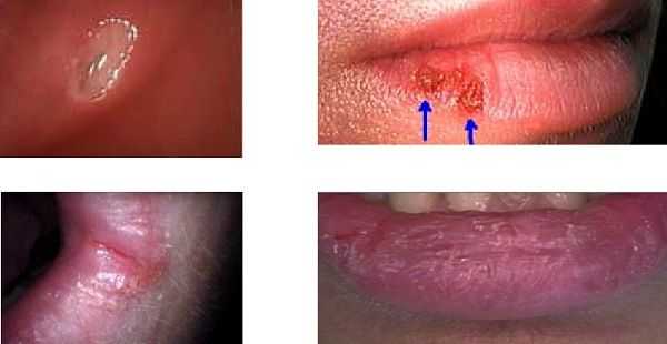
Differential diagnosis: top left – Canker sore (aphthous ulcer) inside the mouth, top right – Herpes labialis (Herpes 1, fever blister, cold sore), bottom left – Angular Cheilitis and bottom right – Chapped Lips. Treatment: An application of an appropriate acidic solution can be most effective in treating a Canker Sore. Anti-viral medication is typically effective in treating an outbreak of Herpes but no current medication will eliminate it from the body. Herpes is highly contagious when it is visible. Angular Cheilitis is an inflammation that occurs at the corners where the upper and lower lips join; it is frequently infected by fungus like Candida (yeast, thrush) that can also infect other parts of the body. It may be contagious. A fungal infection may be treated with Clotrimazole cream. Chapped Lips may be caused by the environment, diet, habits, an allergic reaction, or mouth-dryness associated with other medical conditions or medication. Treatment will depend upon the etiology.
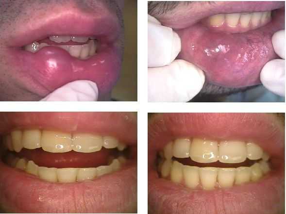
Oral Pathology associated with a lip biting habit: Recurring mucocele. The first image shows a mucocele that was surgically excised by an Oral Surgeon. A biopsy confirmed the clinical impression. The second image was taken one and a half years later when the patient returned with another mucocele in the same location. This was surgically removed. A biopsy again confirmed the diagnosis. The lower two images show before and after incisal adjustment on the sharp lower canine (fang) teeth.
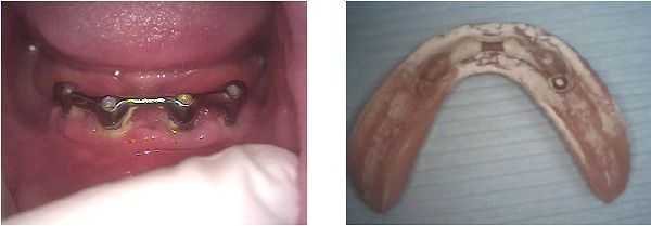
Epulis Fissuratum associated with poor oral hygiene and an ill-fitting implant denture that had a broken clasp. Note the significant plaque around the dental implants in the left photo. Note the calculus (white hardened plaque) on the implant denture. This results from the bacterial infection. Photo #1 of 2
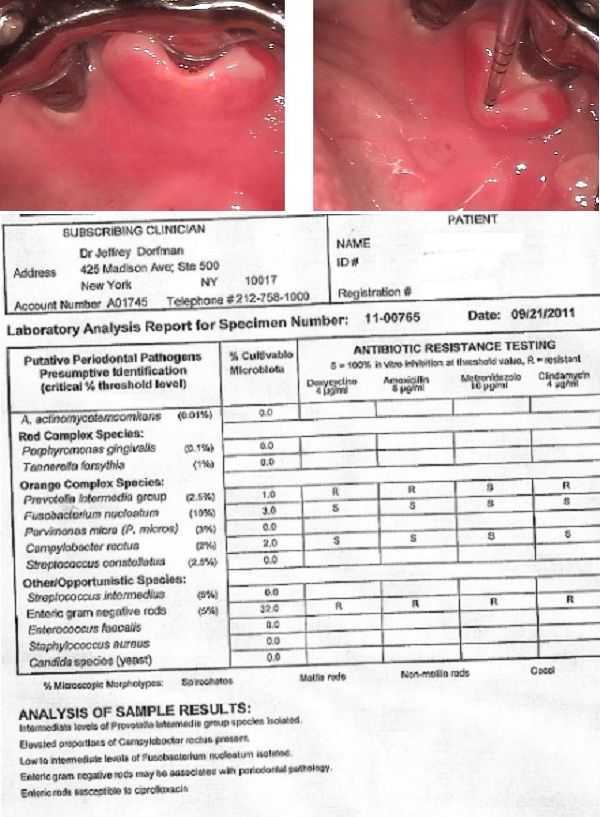
Epulis Fissuratum around 20 year old dental implants in a patient who had not gone to the dentist for over six years. Her implant denture clasps had broken causing the denture to rub against her gums in the presence of a lot of plaque and calculus. These photos were taken after Antibiotic & Initial Periodontal Therapy were completed. The Microbiology Culture revealed the presence of 32% Enteric Gram Negative Rods (5% is normal) that were resistant to Doxycycline, Amoxicillin, Metronidazole and Clindamycin. Antibiotic susceptibility testing revealed this infection would respond to Ciprofloxacin. Note: these two photos were rotated to show the view looking from the tongue. Photo #2 of 2
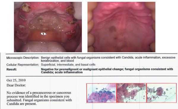
Benign epithelial cells with fungal organisms consistent with Candida (an oral yeast infection) diagnosed in the palate of a middle-aged black female. The melanin deposits in the middle of the palate are the site of the infection and could confuse the diagnosis.
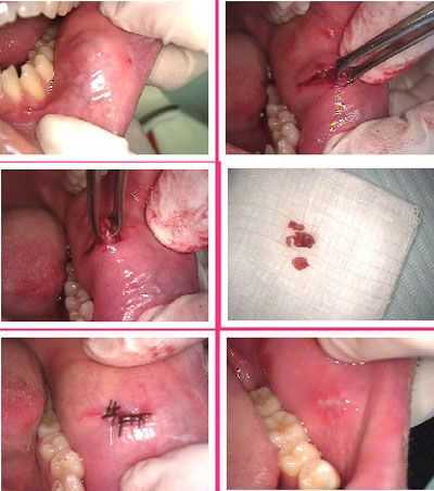
Excision and biopsy of a soft tissue lesion. The oral pathology diagnosis from the Anatomic Pathology Lab of the Columbia University Medical Center was minor salivary gland lobules exhibiting focal, chronic sialadenitis. 1) Pre-operative photo. 2) & 3) Oral surgical removal of the lesion. 4) The soft tissue lesion. 5) Stitches placed. 6) One week post op.
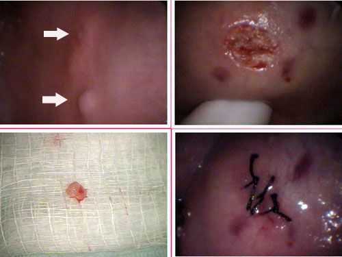
Diagnosis and treatment of an aphthous ulcer – top arrow – and a fibroma – bottom arrow. The next pictures show the excisional biopsy treatment of the fibroma. The fibroma occurred after a cheek bite. Symptoms: aphthous ulcer – pain and fibroma – cheek biting.
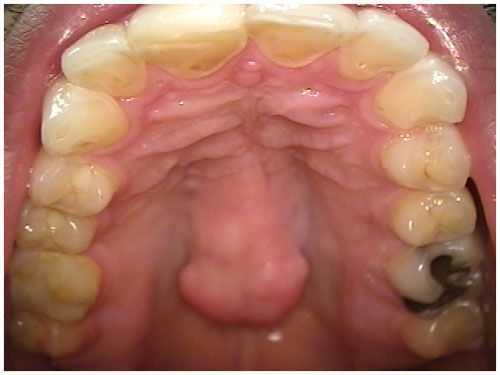
Normal Anatomy. Torus Palatinus or palatal torus. This is normal extra bone in the palate – roof of the mouth -that does NOT change size and there are not any symptoms. No treatment is indicated. The plural is torii. Photo.
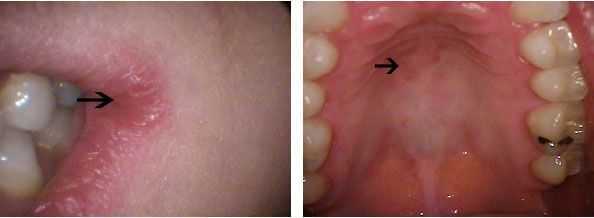
Yeast infections can occur in the mouth in addition to other parts of the body. These pictures show angular cheilitis and a palatal stomatitis due to an oral yeast infection, candida albicans in the mouth. This patient was prescribed Mycelex troches for treatment of the yeast. Symptoms include pain in the corners of the mouth that feel like cuts.
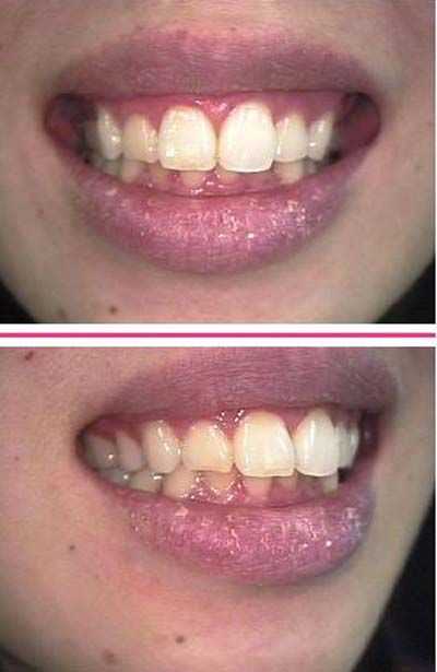
Oral Surgeon examination for a new patient whose symptoms included a complaint of dry lips for several months. Patient states that “it appears to come and go.” No signs of candida, Yeast, or other fungal infections. Intra-orally saliva appeared to be normal in flow and amount. Patient is well hydrated. Suggested treatment is over the counter lip balms for the dryness of the lips. Photos.

An atypical bone pattern was noted on a 46 year old female without symptoms during a routine set of dental x-rays. A Panoramic radiograph also showed something unusual. A subsequent oral surgeon exam revealed no expansion of lingual or buccal cortical plates of the jaw bone. The teeth have not moved for many years though #30 was extracted many years prior. A moderate depression was noted in the jaw bone on the buccal aspect of teeth #’s 29 & 30. The patient exhibited mild pain discomfort in this area upon palpation. A CAT scan confirmed the bone loss and an exploratory surgery treatment was performed. The clinical impression was that of a lateral periodontal cyst. The oral pathology report showed it to be mildly inflamed fibrous tissue. Note the importance of careful evaluation of bone patterns in regular dental x-rays.
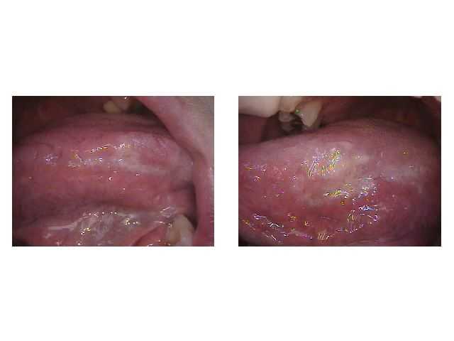
Photos of Bilateral leukoplakia probably hyperkeratosis secondary to a tongue thrusting habit. Geographic tongue also noted. The patient did not have symptoms; no treatment is needed at this time but this patient should be periodically examined for changes.
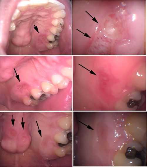
Diagnosis and Treatment Pictures of Necrotizing Sialometaplasia. A 36 year old male presented for a dental prophylaxis cleaning with symptoms of a possible pizza burn near tooth #14 – top photos. Patient was informed of the need for diagnosis and returned two weeks later – middle photos – where the lesion appeared to be healing. He agreed to return four weeks later – bottom photos – where the lesion near tooth #14 appears almost gone but two new lesions appeared on the palatal tori. The patient was informed of the diagnosis and prescribed Kenalog in Orabase for treatment to be used as needed.
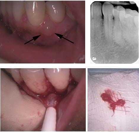
Diagnosis and treatment removal of a Lateral Periodontal Cyst. How to pictures. A 45 year old female patient presents with a firm, non-tender swelling between teeth #’s 21 & 22. There were not associated pain symptoms. Treatment: A gingival incision was made along the necks of teeth #’s 19 – 23 and a full thickness flap was developed. The lesion was identified and initially shelled out using both sharp and blunt dissection. The most lateral aspect of the lesion was in contact with the attached gingiva and could not be completely shelled out. A small portion of the attached gingiva was therefore included in the removal. The lesion was sent for biopsy and the surgical site closed with 4-0 silk sutures. The patient was given antibiotics and pain medication and informed to return for suture removal and reevaluation in one week.
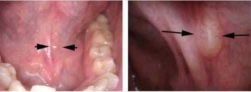
Diagnosis pictures of a ranula. A ranula is a mucocele, or cyst, that forms on the floor of the mouth. It may be associated with a blocked salivary gland. For treatment excision of the ranula is frequently recommended.
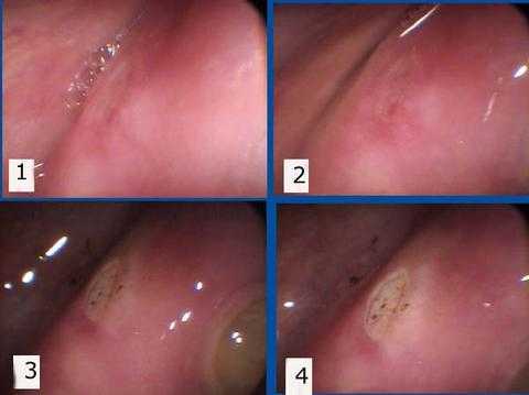
Treatment of a Canker Sore – Aphthous Ulcer. 1) and 2) Pre-op intra oral image of the cheek. 3) and 4) After chemical burn induced by sulfuric acid and sulfonated phenols in an aqueous solution. The most common symptom of a canker sore is significant gum pain.
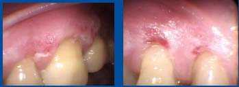
50 year-old female presented with itching symptoms on the buccal gingiva – cheek gums – on the upper left and right. These red lesions and the itching have been present for months. No bone loss, no skin conditions, no ocular conditions. Differential Dx: 1. Non-specific dermatitis – pts mother had a dermatologic condition, or 2. Viral – pt has recurrent herpes. Treatment prescription was – Kenalog. If improvement, it confirms Dx #1. If it gets worse, consider Dx #2. If no change, then change topical steroid. Photos.
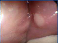
Diagnosis photo of a Fibroma. This was identified during a routine exam and was referred to an Oral Surgeon for diagnosis. Always refer to a specialist for this type of diagnosis. The patient did not have any symptoms.
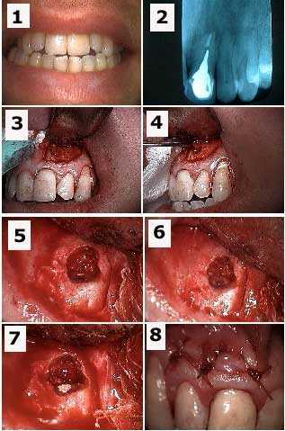
Apicoectomy tooth #9 and retrograde filling to treat a failing root canal for a patient who had tooth pain symptoms. 1) Pre-op photo. 2) Xray of tooth #9 with a cast post and root canal filled with gutta percha to the apex. 3) Initial semi-lunar incision exposed the bone opening fenestration. 4) The dental cyst tissue is pulled through the osteotomy with a forceps. 5) Osseous bone preparation – drilling – exposes the tooth apex with the gutta percha visible. 6) Preparation into the root apex to make room for the retrograde filling. 7) Retrograde filling material (MTA, Mineral Trioxide Aggregate) placed into apex preparation. 8) Sutures.
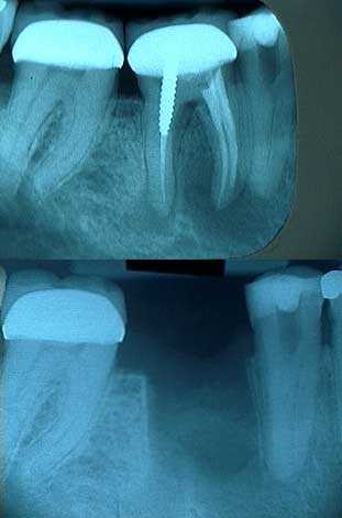
Radiographs of severe periapical pathology around the mesial and distal roots of tooth #30 leading to extraction. This patient had symptoms of severe tooth pain and swollen gums. Initial treatment was tooth extraction.
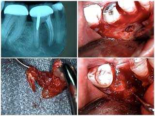
Extraction of tooth #30 due to severe periapical pathology. 1) The radiograph shows severe periapical pathology – tooth infection – around both the mesial and distal roots. 2) Buccal fenestration around the mesial root following a vertical incision and flap. 3) The extracted mesial root with the noted pathology. 4) The extraction site.
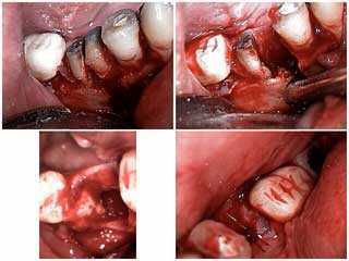
Treatment of tooth pain and swollen gums symptoms. Tooth extraction due to severe periapical pathology – dental infection. 1) Sectioning of the mesial and distal roots. 2) Removal of the mesial root. 3) & 4) The extraction site.
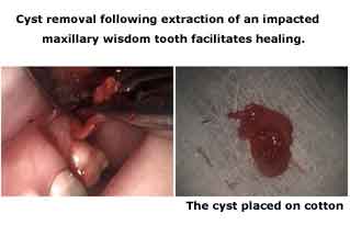
Pictures of a cyst removal following extraction of an impacted maxillary wisdom tooth facilitates wound healing in the tooth socket.
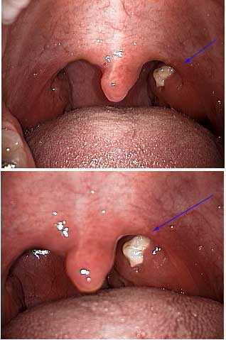
Pictures show a cryptic tonsil with purulence. Patients with a cryptic tonsil may have only mild symptoms.
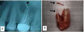
Treatment of a hopeless tooth. 1) Xray shows an upper first molar with gutta percha – root canal filling material – extruded into a large radiolucent area beyond the tooth root apex. 2) Palatal photo of the extracted tooth shows the extent of the gutta percha extrusion. The extra fragment shown is also gutta percha.