Collagen fibers in root dentin cementum and the periodontal tissue and the pulp border

collagen fibers, sharpey fiber bundles,  cementum, connective tissue, SEM  Photomicrographs
Collagen fibers in root dentin, cementum, and the periodontal tissue. Cells, non-collagen matrix and mineral has been removed. Sharpey fiber bundles are seen intermingled between the collagen fibers of the conective tissue in cementum.

Photomicrographs, dental histology pictures.
Copyright Dr. Bo Sogaard-Pedersen, dentistCopenhagen.com

dental photomicrographs, predentin, organic matrix, odontoblast, nerve cell axons, microscopy
Border between pulp and predentin. Cells and non-collagen organic matrix has been removed, as well as mineral. Dentinal canals that harbor odontoblast cell extensions – and nerve cell axons are clearly visible.

Photomicrographs, dental histology pictures.
Copyright Dr. Bo Sogaard-Pedersen, dentistCopenhagen.com

dental photomicrographs, organic matrix, ameloblasts, SEM scanning electron microscope dentistry
During enamel maturation inter-crystalline organic matrix, and water, is re-absorbed by the post-secretory ameloblasts.

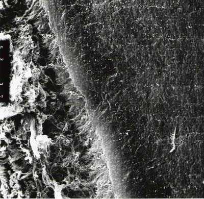
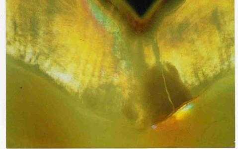
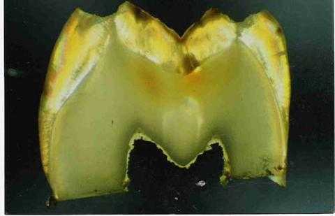
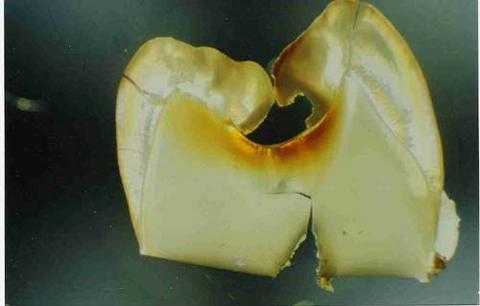
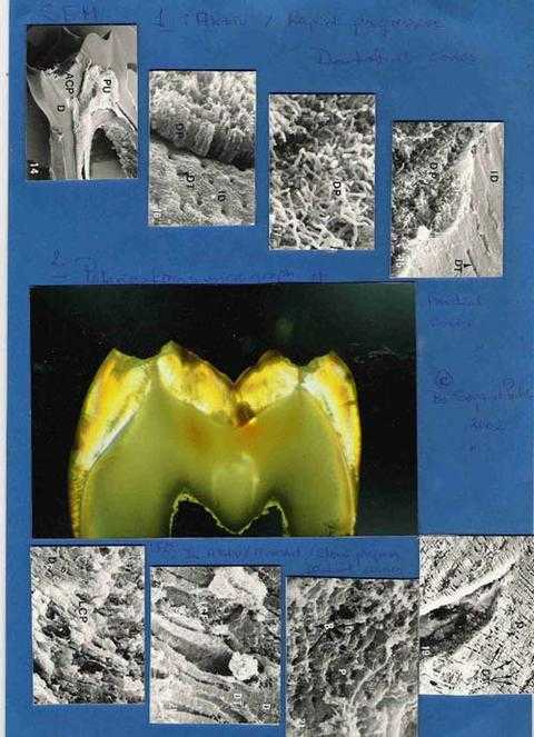
The extensive dehydration of the enamel may induce cracks along its crystals axis, throughout the entire thickness of the enamel, which allows microorganisms to reach the amelo-dentinal junction.

Photomicrographs, dental histology pictures.
Copyright Dr. Bo Sogaard-Pedersen, dentistCopenhagen.com

dental photomicrographs, microbial invasion, inflammation, endotoxins, demineralization, SEM
As a result of microbial invasion of the dental tissue a wide hyper-mineralized “shield”, with the form of a parachute is formed to avoid inflammation or damage to the pulp tissue from endotoxins produced by bacteria; and the dentin immediately adjacent to the micro-organisms establish a dense hypermineralized “sealing” that seems to induce a disruption between enamel and dentin in the circumference of the infected enamel cracks point of contact with dentin.

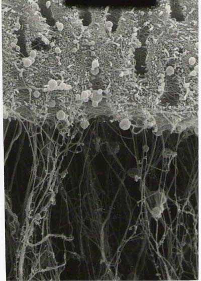
Demineralization of the enamel spread “from the bottom up”, which may indicate that anaerobic micro-organisms at this stage are responsible for the increased solubility of the enamel crystals and extraction of mineral from these.

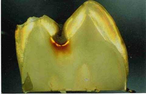
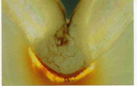
The demineralized enamel forms a pyramid-like area with its basis next to the dentin.

Photomicrographs, dental histology pictures.
Copyright Dr. Bo Sogaard-Pedersen, dentistCopenhagen.com

dental photomicrographs, SEM,  demineralization, amelo-dentinal border, microscope
The demineralization of enamel spread along the amelo-dentinal border, as do the detachment of the enamel from dentin. The crystalline structure of the enamel is transformed into an amorph / extensively dissolved state around the crack, and beyond.

Photomicrographs, dental histology pictures.
Copyright Dr. Bo Sogaard-Pedersen, dentistCopenhagen.com

dental photomicrographs, dissolved enamel, proteolysis, SEM scanning  electron microscope
Dentin starts getting a brown discoloration underneath the amorph / dissolved enamel, and is partly demineralized in this zone. Although it has not yet suffered proteolysis of the organic crystallization focus, – or collagen fibers.

Photomicrographs, dental histology pictures.
Copyright Dr. Bo Sogaard-Pedersen, dentistCopenhagen.com

dental photomicrographs, dissolved enamel, remineralization, SEM scanning electron microscope
A cavity in the enamel is established as the amorph / extensively dissolved enamel is broken down. The dentin still retains its ability for remineralization, although mineral is lost to some depth.

Photomicrographs, dental histology pictures.
Copyright Dr. Bo Sogaard-Pedersen, dentistCopenhagen.com

dental photomicrographs, SEM, Scanning Electron Micrographs, proteolytic, foci  liquefactions
A sample of Scanning Electron Micrographs of acute non-proteolytic dentinal caries: “bioleaching”, and carious dentin under proteolytic destruction: gross invasion of dentinal tubules are seen, along with liquefactions foci, split dentinal fissures, and a purulent pulp chamber where the connective tissue and odontoblasts have been destroyed.

Photomicrographs, dental histology pictures.
Copyright Dr. Bo Sogaard-Pedersen, dentistCopenhagen.com