Two-unit bridge with an occlusal rest seats onto the rest preparation in the mesial of the second molar crown. This type of semi precision attachment eliminates the need for removing the second molar crown. The premolar did receive root canal therapy following healing of the extraction site.
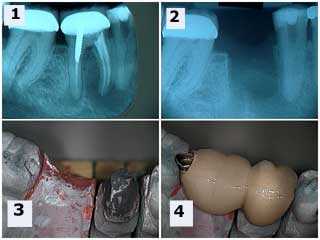
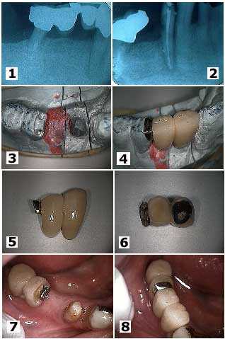
Cervical decay and root canal infection around a lower right first premolar. 1) A large radiolucency is seen. Note the distal abutment is the distal root of a first molar following hemisection. 2) Following root canal therapy. The cervical decay can now be seen more easily. 3) The working model showing the preparation of the mesial part of the distal two teeth bridge. This semi precision attachment will allow rigid connection to the new anterior bridge being made. A “T” shaped female preparation should be considered when possible. 4) The new bridge section in place on the working model. 5) & 6) Two views of the new bridge showing the distal attachment. 7) An intraoral view which mirrors the working model view seen in 3). 8) The final result. This patient was content to see the gold occlusal rest. Otherwise, it could have been hidden under porcelain.
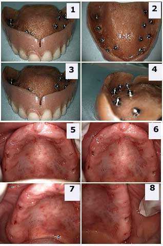
Intramucosal Insert Implants are a simple and effective means to retain a maxillary complete or partial denture via the implant attachment to the soft tissue gums. These mushroom-shaped titanium implants are embedded into the acrylic underside of a denture. Special burs create soft tissue receptor sites in the palatal tissue that retains these implants. The technique is described in the Dental Textbook section located above.
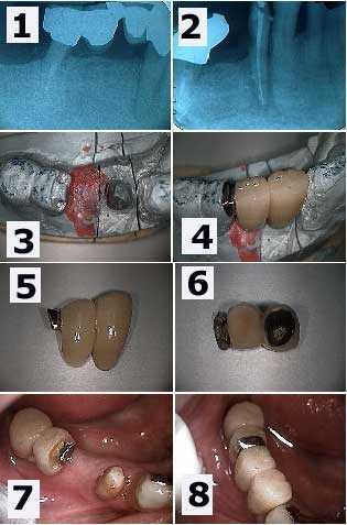
These pictures show to to use a semi precision attachment to connect one porcelain metal teeth bridge to another porcelain metal teeth bridge in the middle of a bridge pontic. 1) This elderly patient developed distal root caries under a bridge and a root canal infection on a lower first premolar. Note that the distal molar had previously been hemisected. 2) Following bridge section removal and root canal therapy. 3) – 4) Two and a half unit bridge fabricated and seated on the working model. 5) – 6) Two views of the two and a half unit bridge. The distal half unit has been fabricated with a female semi-precision attachment. 7) Intra-oral view. Note how the metal framework of the pontic was prepared into a “T” shape to help lock in the bridge upon cementation. 8) The final bridge.