Before and after photos on apicoectomy is root canal oral surgery performed in our Extraction Oral Surgery office.

Apicoectomy is root canal oral surgery that is needed when traditional root canal therapy has failed and cannot be retreated through the root canal of the dental crown.
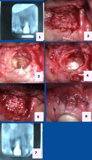
Apicoectomy tooth #9. 1) Pre-op x-ray. 2) Isolation of the apical area. 3) Exposure of the root apex. 4) Placement of MTA (mineral trioxide aggregate). 5) Placement of Bioplant HTR (hard tissue replacement). 6) 6-0 Vicryl sutures. 7) Post-op x-ray.

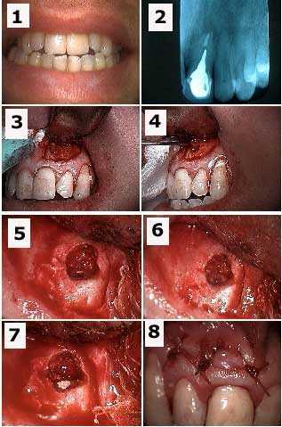
Apicoectomy tooth #9 and retrograde filling to treat a failing root canal. 1) Pre-op photo. 2) X-ray of tooth #9 with a cast post and root canal filled with gutta percha to the apex. 3) Initial semi-lunar incision exposes the bony fenestration. 4) Cystic tissue is being pulled through the osteotomy with a forceps. 5) Osseous preparation exposes the tooth apex with the gutta percha visible. 6) Preparation into the root apex to make room for the retrograde filling. 7) Retrograde filling material (MTA, Mineral Trioxide Aggregate) placed into apex preparation. 8) Sutures.

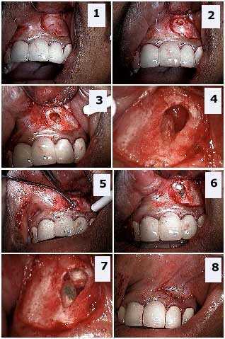
Apicoectomy and retrograde filling. 1) Upper left lateral incisor shown with tissue reflected. 2) Initial penetration through cortical bone. 3) Tooth apex located. 4) Tooth root after sectioning off the apex. 5) Application of Mineral Trioxide Aggregate (MTA) retrograde filling material. 6) – 7) MTA in place. 8) Closure.