Before and after photos on crowded impacted teeth need extraction performed in our Extraction Oral Surgery office.
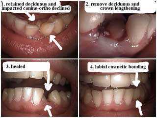
The labial surface was bonded to close the tooth gap space after tooth extraction of the impacted canine tooth, and healing, of the deciduous tooth.
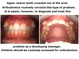
Impacted teeth – the maxillary canines – crowded out of the arch.
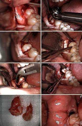
Extraction of a lower right partial bony impacted wisdom tooth. 1) Soft tissue reflection. 2) & 3) Sectioning of the molar. 4) An elevator is being used to split the molar. 5) & 6) Lower forceps and an elevator are used to extract the split roots. 7) The extracted tooth in two sections. 8) Closure of the surgical site.
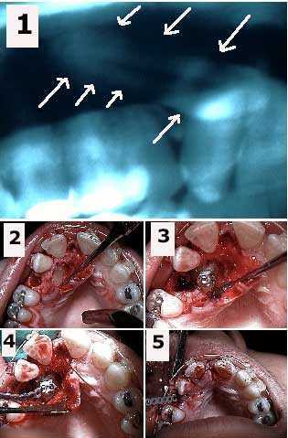
Surgical exposure of a palatally impacted upper canine tooth and placement of an orthodontic bracket and elastic power chain. 1) Panorex showing the horizontal impaction of the upper right canine. 2) Initial exposure of the impacted canine and extraction site after removal of an over-retained deciduous tooth. 3) Orthodontic bracket cemented to the canine. 4) Elastic orthodontic power chain attached to the bracket. 5) Emergence of power chain through the extraction site after suturing. The power chain will be attached to the arch wire to exert a pulling force on the canine to bring it into the area of the present extraction site. The canine will assume a normal position in the arch in 18-24 months.
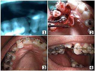
Orthodontic eruption of a palatally impacted canine tooth. Eleven months after the surgical exposure of the canine. Follow up pictures.
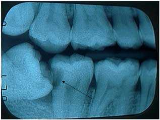
Rationale for extraction of impacted wisdom teeth. The lower wisdom tooth is on an angle and leaning – impacted – against the second molar. This created a huge cavity – tooth decay – in the second molar that will probably require root canal therapy and a dental crown to repair. The patient did not feel tooth pain.