Photos on oral surgery cases include tongue tied and accidents created in our Extraction Oral Surgery office.
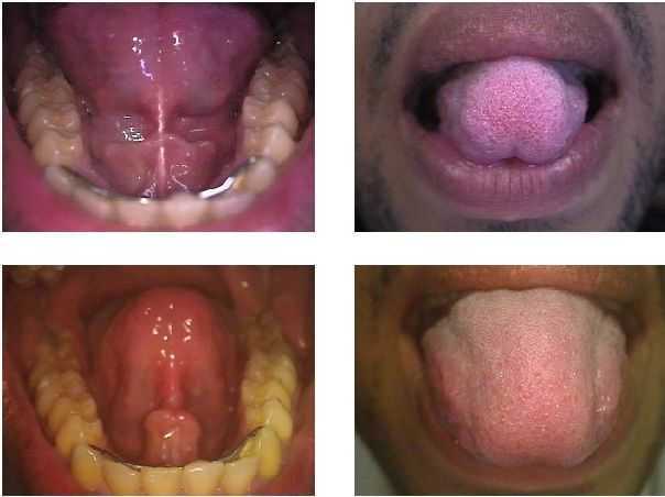
Treatment of a tongue-tied patient. The top photos were taken before treatment and the bottom photos were taken following a frenulectomy. A frenulectomy is a conservative procedure, typically performed by an Oral Surgeon or Periodontist, that involves cutting the lingual frenum to allow more movement of the tongue.
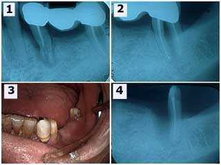
Oral surgery treatment following accidental trauma to a teeth bridge. 1) The initial xray showing the three teeth bridge and severe damage to the second premolar tooth. 2) The second molar tooth has a furcation involvement and thickening of the periodontal ligament space – pdl space – around the mesial root. The second premolar tooth and the mesial root of the second molar were extracted. 3) Photo following wound healing. The patient declined dental implants and wanted a new longer span fixed dental bridge. 4) Radiographic healing of the distal root of the second molar after six weeks. Sometimes – even in this age of dental implants – there is still value in saving individual roots of molar teeth.
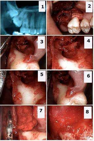
Extraction of two impacted teeth by an Oral and Maxillofacial Surgeon. How to pictures: 1) Xray radiograph shows the double impaction of teeth # 1 and 2. Photos 2) – 6) Different views showing the large osseous defect in bone and the significant exposure of the distal furcation of the first molar tooth. 7) – 8) The bone defect is packed with a freeze-dried bone graft and gelfoam.
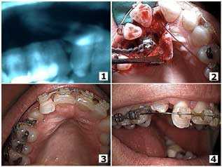
An Oral and Maxillofacial Surgeon and Orthodontist work together on the treatment of the orthodontic eruption of a palatally-impacted canine tooth. 1) Starting x-ray. 2) Starting photo. 3) and 4) Pictures show eleven months after the surgical exposure of the canine tooth.