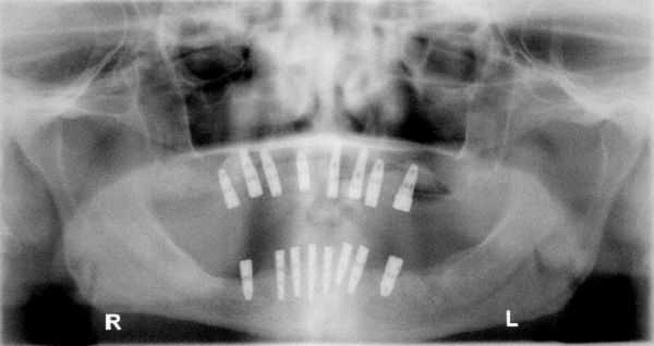
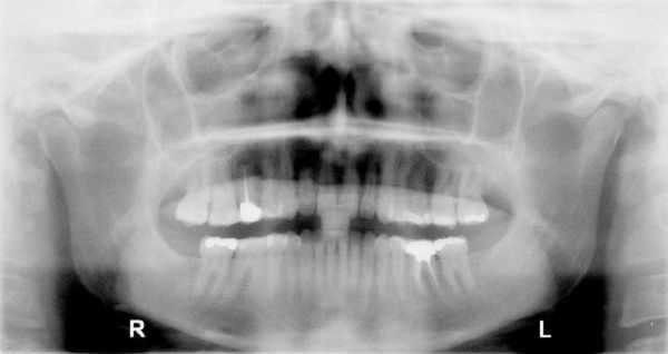
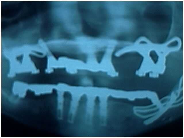
Photos and X-rays on panoramic x-rays are full mouth dental radiographs created in our Extraction Oral Surgery office.


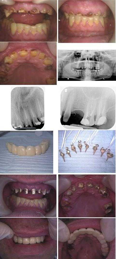
Oral Rehabilitation Dental Reconstruction of a dental fear phobia patient who is a Wall Street executive. Focus on dental radiographs x-rays: a panoramic x-ray and two periapical xrays. Total treatment time was about nine hours in two visits during one business week: Monday (four hours) and Friday (five hours). This patient hid his dental condition for over a decade by never showing his smile.
In the first row of photos, note that the dental bite occlusion was over-closed due to a prior history of an eating disorder, now controlled, and a current teeth grinding habit bruxism.
An Oral Surgery consultation with panoramic xray (second row) was performed prior to treatment to evaluate opening the bite.
The third row shows x-rays taken after the Endodontist completed eight root canals on the first day on tooth #’s: 6 – 11 and 14, 15.
The fourth row shows the lab-processed 11 unit (11 tooth) temporary dental bridge and the cast gold post and cores with Kaitlyn loops for the root canal treated teeth.
The fifth row shows the cast gold post and cores cemented.
The sixth row shows the lab-processed provisional temporary teeth bridge in place after the Oral Surgeon extracted tooth #’s: 3 – 5 and 12, crown lengthening gum surgery was performed on tooth #’s: 6 – 11, and a distal wedge was performed on #15.
The patient will have a final porcelain metal dental bridge made after the gums heal. Dental implants may also be placed in the upper right posterior. A bite plate night guard is also necessary to try to mitigate the force of teeth grinding clenching. Referral for pharmacological management of anxiety is also worthwhile.
