Before and after photos on the maxillary and mandibular jaw bone and alveolar ridge performed in our Extraction Oral Surgery office.
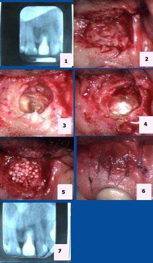
Apicoectomy Pictures of tooth #9. 1) Pre-op x-ray. 2) Isolation of the apical area. 3) Exposure of the root apex. 4) Placement of MTA (mineral trioxide aggregate). 5) Placement of Bioplant HTR (hard tissue replacement). 6) 6-0 Vicryl sutures. 7) Post-op x-ray.
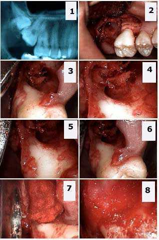
Extraction of a double impaction in the upper jaw, maxilla: upper right third (wisdom) and second molars. 1) x-ray, Radiograph, showing the double impaction. 2) – 6) Different picture views showing the large osseous defect and the significant exposure of the distal furcation of the first molar. 7) – 8) Packing the defect with freeze-dried bone and gelfoam.