Before and after photos on X-rays and digital radiographs in oral surgery performed in our Extraction Oral Surgery office.

Radiographic case study. An atypical bone pattern was noted on a 46 year old female during a routine set of dental x-rays. A Panoramic xray radiograph also showed something unusual. A subsequent Oral Surgeon exam revealed no expansion of lingual or buccal plates. The teeth have not moved for many years though #30 was extracted many years prior. A moderate depression was noted on the buccal aspect of teeth #’s 29 & 30. The patient exhibited mild discomfort in this area upon palpation. A CAT scan confirmed the bone loss and an exploratory oral surgery was performed. The clinical impression was that of a lateral periodontal cyst. The pathology report showed it to be mildly inflamed fibrous tissue. Note the importance of careful evaluation of bone patterns in a routine dental x-ray series.
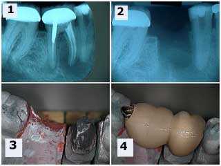
One periapical x-ray radiograph taken before a molar tooth extraction and one periapical xray taken after the tooth was extracted. A two teeth bridge with an occlusal rest seat on the rest preparation in the mesial of the second molar dental crown. This eliminates the need for removing the second molar tooth crown. The premolar did receive root canal treatment following wound healing of the tooth extraction oral surgery.
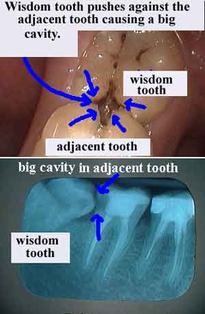
One photo and one dental radiograph x-ray explain the reason for Wisdom Tooth Extraction oral surgery. In the picture the wisdom tooth can be seen growing at an angle directly into the adjacent tooth causing a tooth cavity. In the radiographic xray view the extent of the damage from the dental caries may be seen.
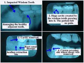
This x-ray radiographic series shows how an impacted wisdom tooth damages the healthy adjacent tooth. The second xray was taken after tooth extraction oral surgery and shows that a tooth cavity – dental caries – was created in the adjacent second molar by the wisdom tooth growing into it. The patient did not have any symptoms of tooth pain.
Treatment: Root canal and crown (the crown covers the site where decay was removed.
Treatment: Root canal and crown (the crown covers the site where decay was removed.
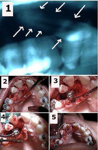
Panoramic radiograph x-ray of the oral surgery exposure of a palatally impacted upper canine tooth and placement of an orthodontic bracket and elastic power chain. 1) Panoramic xray shows the horizontal tooth impaction of the upper right canine. 2) Initial oral surgical exposure of the impacted canine and the tooth extraction site after removal of an over-retained deciduous tooth. 3) The Oral Surgeon placed dental cement on the orthodontic bracket to attach it to the canine tooth. 4) Elastic orthodontic power chain attached to the bracket. 5) Emergence of power chain through the extraction site after suturing. The power chain will be attached to the arch wire to exert a pulling force on the canine to bring it into the area of the present extraction site. The canine will assume a normal position in the arch in 18-24 months.
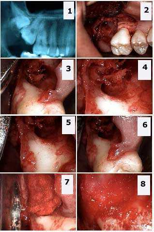
Preoperative Oral Surgery panoramic radiograph x-ray. Tooth extraction of a double tooth impaction: the upper right wisdom third molar and the upper right second molar. 1) This radiograph xray shows the double impaction. 2) – 6) Different pictures show the large osseous bone defect and the significant exposure of the distal furcation of the first molar. 7) – 8) These photos show the Oral Surgeon packing of the osseous defect with freeze-dried bone and Gelfoam.
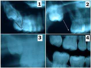
A panoramic x-ray radiographic series shows the sequence of wisdom tooth eruption after a mistaken extraction of a second molar. This patient is a 27 year-old female who had recently moved to New York from Seattle. She brought prior xrays radiographs and an interesting story involving oral surgery in Seattle that I thought was worth relating. 1) In Seattle the patient was referred to an oral surgeon for the extraction of #1 but #2 was erroneously extracted instead. 2) Radiograph xray after tooth extraction of #2 shows that #1 was still present. 3) This new, less clear Panoramic x-ray was taken a year later (still in Seattle) but the wisdom tooth #1 can be seen moving down into the second molar position on its own. 4) This bitewing x-ray radiograph was the first of the series taken in our office. The wisdom tooth #1 has almost fully erupted on its own after about two and a half years. PS – the patient was billed for and did pay for the extraction of the wrong tooth!
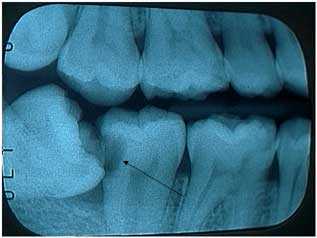
This bitewing radiograph x-ray explains why a wisdom tooth should be extracted. The lower wisdom tooth is on an angle and leaning against the second molar. This has caused a huge dental cavity – tooth decay – in the second molar that will probably require root canal treatment and a dental crown to repair. The patient did not feel tooth pain symptoms in this tooth. This patient scheduled for oral surgery.
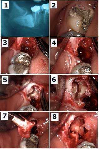
The Oral Surgery extraction of a horizontally-impacted lower wisdom tooth and significant dental caries tooth decay in the adjacent second molar. This patient had severe tooth pain symptoms and swollen gums. Xray and Pictures. 1) Pre-op panoramic radiograph x-ray. 2) Tooth #31 had a temporary dental filling in it. 3) – 4) These photos show after the removal of the dental crown of #32 showing the residual empty tooth socket. 5) – 6) The Oral Surgery raised a minimal gingival gum flap to show exposure of the roots of #32 with sectioning. 7) Photo shows removal of the first tooth root. 8) Second root brought forward in socket and ready for removal.