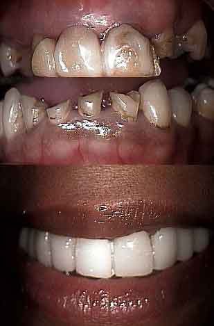
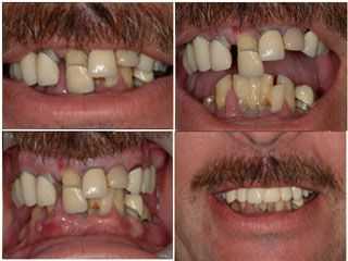
Before and after photos on dental reconstruction for teeth grinding or fear performed in our Smile Makeover office.
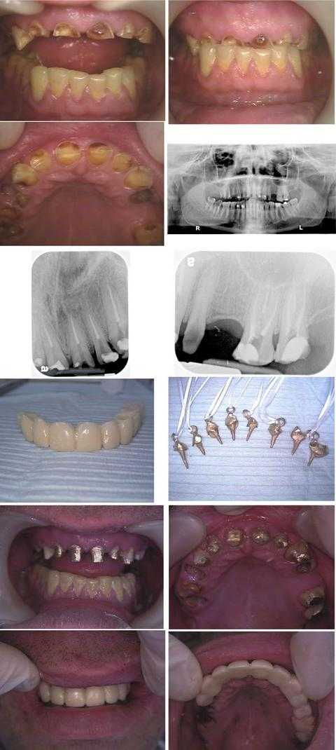
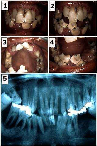
How to pictures: Oral rehabilitation dental reconstruction smile makeover. This Wall Street executive suffered from dental fear phobia. Total treatment time was about nine hours in two visits during one business week: Monday (four hours) and Friday (five hours). This patient hid his dental condition for over a ten years by hiding his smile.
In the first row of photos, note that the teeth bite occlusion was over-closed due to a prior history of a bulimia eating disorder, now controlled, and a current teeth grinding habit.
An Oral Surgery consultation with panoramic x-ray (second row) was performed prior to treatment to evaluate opening the dental bite.
The third row shows x-rays taken after the Endodontist completed eight root canal treatments on the first day on teeth #’s: 6 – 11 and 14, 15.
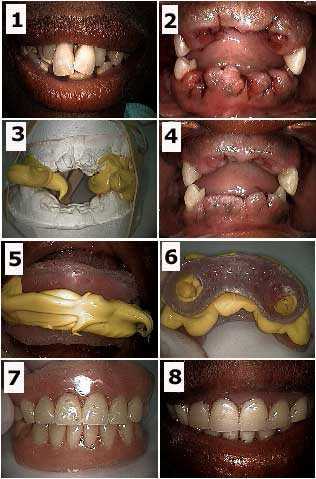
The fourth row shows the dental lab-processed 11 teeth provisional temporary dental bridge and the cast gold post and cores (with Kaitlyn loops) for the root canal treated teeth.
The fifth row shows the cast gold post and cores after being placed with dental cement.
The sixth row shows the lab-processed temporary teeth bridge in place after the Oral Surgeon extracted teeth #’s: 3 – 5 and 12, crown lengthening gum surgery was performed on teeth #’s: 6 – 11, and a distal wedge was performed on #15.
The patient will have a final porcelain-metal teeth bridge – attached dental crowns – made after the gums heal. Dental implants may also be placed in the upper right posterior. A bite plate night guard is also necessary to try to mitigate the force of teeth clenching grinding. Referral for pharmacological management of anxiety is also worthwhile.
Initial full mouth dental reconstruction on a dental phobia patient. Before and after pictures. Note that the patient had been wearing a very old, broken temporary teeth bridge; she had been recementing it herself daily for years because she was afraid to visit a dentist. The after photo shows a new lab processed temporary dental bridge placed later that same day. It took this dental fear anxiety patient six visits before she was comfortable enough to begin dentistry. We patiently waited until she was emotionally ready to begin her oral rehabilitation smile makeover treatment.
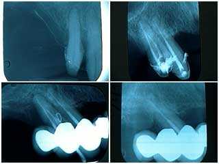
Radiographs x-rays of a failing distal tooth abutment in a one year old, ten teeth fixed porcelain crown dental reconstruction. How to repair xrays. 1) This pre-operative xray show the initial teeth abutment preparation. 2) This x-ray was taken after root canal therapy, root planing and open periodontal gum debridement. Note this premolar tooth had multiple roots; multi-rooted maxillary premolar teeth typically have a more guarded prognosis when performing oral rehabilitation. 3) Gutta percha placed into a periodontal gum abscess one year later. 4) Following root planing, targeted antibiotics and tooth extraction. The roots of this extracted tooth were fused. It is suggested that dental rehabilitation smile makeover patients are treated, not cured, and that they must be closely supervised. Patients need to be acutely aware of the need for frequent dental hygiene recall visits with careful examination by both the dental hygienist and dentist.
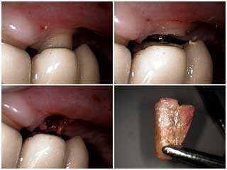
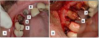
These pictures teach the technique method for removal of a failing distal abutment tooth under a long span fixed, porcelain fused to metal dental bridge oral rehabilitation. 1) Photo of the upper first premolar tooth with periodontal probing to the tooth root apex. The Periodontist determined this tooth was not amenable to periodontal gum treatment. 2) Horizontal sectioning through the tooth to separate it from the dental reconstruction teeth bridge. The dentist should remove enough of the remaining tooth root to be able to elevate it while it is under the bridge. 3) Tooth socket after tooth removal. 4) Extracted tooth root. The gingival surface of the newly created pontic could be bonded after primary healing has occurred.
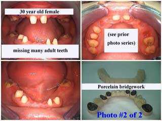
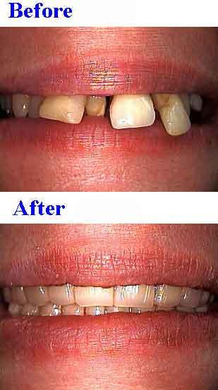
Oral Rehabilitation with porcelain dental crowns for a partial anodontia patient. This 30 year old female had many congenitally missing adult teeth, retained baby teeth and a closed teeth bite occlusion. Before and after photos of a dental reconstruction. Photo #1 of 2
Oral Rehabilitation with porcelain dental crowns for a partial anodontia patient. This 30 year old female had many congenitally missing adult teeth, retained baby teeth and a closed teeth bite occlusion. How to treatment pictures. Photo #2 of 2.
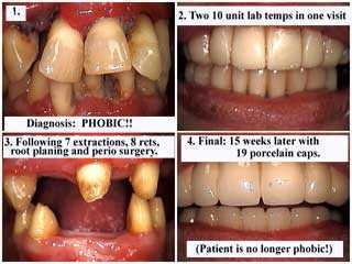
Dental Rehabilitation Reconstruction of a dental phobia fear patient. Before and after pictures. First treat the dental fear and then fixing the teeth becomes easy. The patient was restored to a first premolar occlusion.
Dental Treatment: 7 teeth extractions, 8 root canal treatments and composite resin cores, full mouth scaling and root planing and then periodontal gum surgery, 19 units of bridgework – 19 total dental crowns in the upper and lower jaws – over 8 abutment teeth. The patient is no longer phobic and has since retired in Florida. She flies up for the day once every three months for dental hygiene visits and checkups. These teeth bridges are now over ten years old.
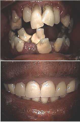
Dental Reconstruction of a dental anxiety patient who was very scared of dentists. The Center for Special Dentistry does treat a lot of people who are afraid and need oral rehabilitation. Treat the fear first; take time to understand the emotional needs of a patient and then the dentistry becomes easy.
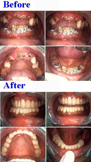
This patient received 20 provisional temporary caps crowns, 14 root canal treatments and 6 teeth extractions in one visit with a carefully planned choreography of cosmetic dentists, specialists and lab technicians.
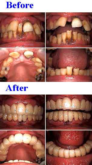
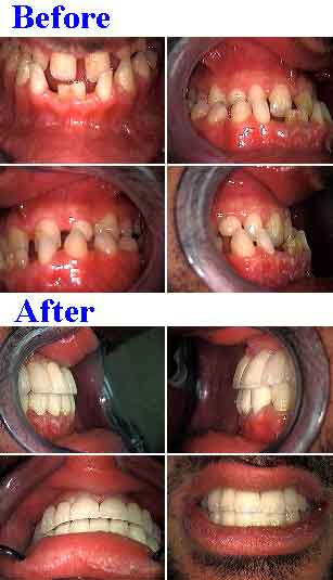
Smile makeover of an underbite – lower jaw in front of the upper jaw or class 3 – and retained baby teeth. Two 14 teeth porcelain metal dental bridges were used to increase the vertical dimension of occlusion and “jump” the cross bite. Dental reconstruction for a 21 year old male who had a prognathic lower jaw and refused orthognathic options. Treatment time – 16 days. Photo #1 of 2.
Smile makeover of an underbite – lower jaw in front of the upper jaw or class 3 – and retained baby teeth. Close up pictures. Two 14 teeth porcelain metal dental bridges – one for each jaw – were used to increase the vertical dimension of occlusion and “jump” the cross bite. Dental reconstruction for a 21 year old male who had a prognathic lower jaw and refused orthognathic options. Treatment time – 16 days. Photo #2 of 2.
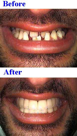
Full mouth Oral Rehabilitation for a 40 year old, married owner of a jewelry company who suffered from dental fear. This man built a successful business by telephone to avoid meeting clients in person. This was a typical New Years resolution smile makeover case. Treatment included dental laboratory processed provisional temporary crowns for the upper – maxilla – and lower -mandible – jaws. Treatment time – four days. More dentistry is still needed.
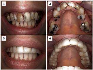
It’s NOT about the teeth! It’s about the human being attached to them. Full mouth dental reconstruction of a patient who was scared of dentistry and afraid of the dentist. This before and after photo of a very intelligent and very successful businesswoman took one visit. The treatment provided included ten upper jaw and ten lower jaw temporary dental crowns, root canal treatment for 14 teeth and 6 teeth extractions.
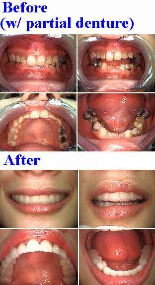
Full mouth reconstruction, oral rehabilitation of a 36 year old female dental fear phobia patient. Before and after pictures – two visits. Photo #1 of 4.
Full mouth dental reconstruction of a phobic 36 year-old female. Initial visit. Initial pictures and panoramic radiograph x-ray. It is important to determine what teeth, if any, may be saved at least temporarily. It is easier for an Oral Rehabilitation patient to emotionally adjust to a temporary prosthesis that has at least some amount of retention provided by natural teeth abutments. The teeth chosen were #6, 11, 22 and 28. The decision to fabricate a removable immediate partial denture, rather than a fixed lab-processed fixed temporary dental bridge, was determined by the particular weakness of tooth #28. The patient was informed that the immediate prosthesis was to be used for the healing phase and that the four remaining teeth abutments, particularly #28, might be subsequently extracted. Photo #2 of 4.
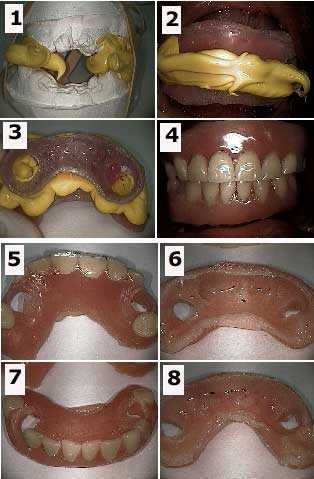
Dental rehabilitation of a phobic 36 year-old female. How to pictures. 1) Initial before photo. 2) Following teeth extraction of all but the four remaining abutments. 3) Working models and teeth bite made right after the teeth extractions. This will serve for the fabrication of the immediate denture. 4) Three days following the extractions. 5) Intra oral photo of the bite rims and bite material. 6) Extra oral photo of the bite rims and bite material. The patient did not want any wire clasps to be used to retain the removable prosthesis. 7) Extra oral after photo of the immediate dentures. 8) Intra oral after photo of the immediate dentures. A soft reline material was used on the gingival gum side of these dentures. This soft reline material was allowed to flow into the circular openings (see photo #6) in the acrylic denture as it was seated. This soft reline material provided retention instead of using clasps. This smile makeover took two visits over three days. Photo #3 of 4.
Full mouth oral rehabilitation of a 36 year-old female dental anxiety fear patient. How to pictures. 1) Working models and dental bite made right after the teeth extractions. This will serve for the fabrication of the immediate denture. 2) Intra oral photo of the bite rims and bite material. 3) Extra oral photo of the bite rims and bite material. The patient did not want any wire clasps to be used to retain the prosthesis. 4) Intra oral photo of the immediate dentures. A soft reline material was used on the gingival side of these dentures. This soft reline material was allowed to flow into the circular openings in the acrylic denture as it was seated. This soft reline material provided retention instead of using clasps. 5) & 6) Images of the upper denture with soft reline material visible. 7) & 8) Photos of the lower denture with soft reline material visible. Photo #4 of 4.
Upper Jaw dental reconstruction in a patient who was scared of the dentist. The patient was a pretty, 33 year-old female. Treatment included porcelain fused to metal dental crown & bridge, root canal therapy on all teeth abutments, root tip extractions, and facial teeth bonding on both upper lateral incisors. The upper anterior root canal and dental crowns were first completed to show her how pretty her teeth could look and that it could be accomplished quickly and painlessly. Extractions of the hopeless teeth were all completed at one time and initiated early to allow time for healing before fabrication of the final porcelain teeth crowns bridgework. Then root canal therapy was performed on the posterior abutments before their preparation for bridgework. The patient experienced minimal post-operative tooth pain. Multi-specialty oral rehabilitation involving a Cosmetic Dentist, Oral Surgeon, Endodontist and Dental Laboratory Technician.
Teeth Reconstruction performed by a Columbia University dental student at The Center for Special Dentistry. Before and After pictures. The student, Jared Bowyer, is part of a unique honors program at Columbia University Dental School wherein dental student interns learn how to perform full mouth Oral Rehabilitation in our office.
Selective teeth extraction during an upper dental reconstruction. The initial treatment plan considered saving teeth #’s 6, 7 and 9 while extracting #’s 5 and 8. During the oral surgery, #5 was considered more stable than #6 so #5 was retained and #6 was extracted. The retained teeth will be used to support a fixed temporary dental bridge until subsequent dental implants are placed and heal.

