Before and after photos on smile diagnosis needs photos of a smiling face performed in our Smile Makeover office.
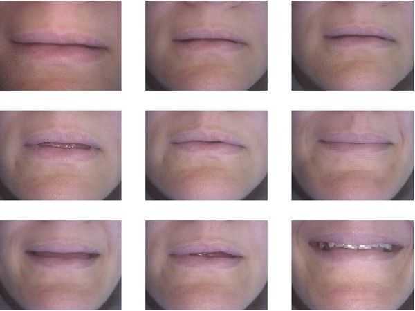
Smile diagnosis. You cannot see this patients teeth when she speaks. This is because she exhibits a closed bite. Treatment options include orthognathic surgery or cosmetic dentistry. Cosmetic dentistry could include using either porcelain onlays or crowns on the maxillary (upper) posterior teeth and porcelain veneers on the maxillary anterior teeth.
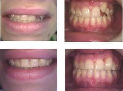
Cosmetic Dentistry with one tooth bonding and one porcelain dental veneer. Treatment time: 2 visits. The patient refused orthodontic teeth braces treatment. The tooth veneer had pink porcelain around the gingival margin gums to hide its relative height.
Lip Line Analysis focuses on a patient’s face and smile while relaxed, speaking and when smiling. It provides valuable information before beginning cosmetic dentistry and/or oral rehabilitation.
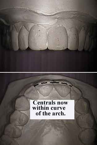
Analysis of a patient’s smile with study models. It is usually much easier to examine a cosmetic dentistry problem outside of the mouth. This patient originally had a constricted narrow upper jaw that was treated with fixed teeth braces for six months. It may be difficult for a patient to see what you want them to see while they are smiling.
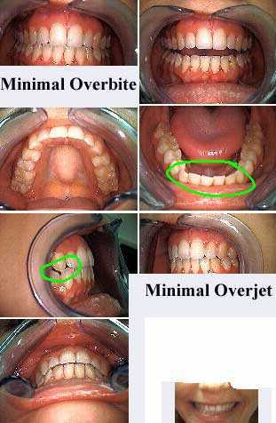
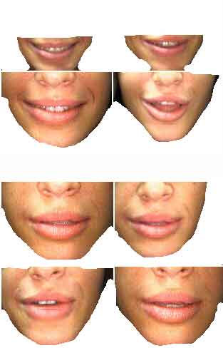
Smile Analysis & Facial Profile. Photographic Survey Only. This supermodel demonstrates only 1 mm of both overbite and overjet. This results in aesthetically beautiful smiles. Overbite is the vertical overlap of upper teeth over lower teeth. Overjet is the horizontal overlap of upper teeth over lower teeth.
Cosmetic dentistry using dental bonding can quickly improve the face smile of a fashion model.
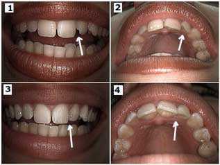
In this case bonding was placed to make a rotated, upper lateral incisor tooth appear straight. Before and after pictures. 1) Pre-operative front photo. 2) Pre-operative occlusal photo. Note how the tooth is rotated. 3) Post-operative front photo. 4) Post-operative occlusal photo. Note how tooth bonding was added to the mesio-palato-incisal angle of the lateral tooth. The other lateral will be treated next.
In this case bonding was placed to make a rotated, upper lateral incisor tooth appear straight. Before and after pictures. 1) Pre-operative front photo. 2) Pre-operative occlusal photo. Note how the tooth is rotated. 3) Post-operative front photo. 4) Post-operative occlusal photo. Note how tooth bonding was added to the mesio-palato-incisal angle of the lateral tooth. The other lateral will be treated next.