Before and after X-rays of a smile makeover or oral rehabilitation performed in our Smile Makeover office.
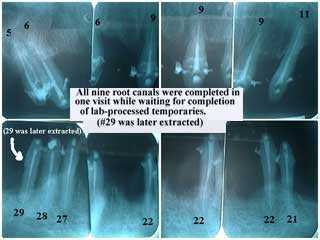
Radiographic series shows eight periapical x-rays of teeth abutments during a dental reconstruction smile makeover. These xrays were taken after the Endodontist completed root canal treatment on nine teeth while the Cosmetic Dentist and Dental Lab Technician completed fabrication of the dental lab-processed provisional temporary dental crowns. In each radiograph x-ray note endodontic obturation to the end of each tooth root apex. #29 was later extracted for periodontal reasons.
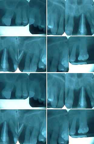
Preoperative radiographs x-rays for a dental reconstruction smile makeover. How to begin full mouth oral rehabilitation. First learn the non-dental origin of the problem and think how to treat it. Study the patient’s face and perform a clinical exam. Compare the clinical exam to the radiographic xray diagnosis. Sometimes a panoramic x-ray may be taken if either the patient will need oral surgery like teeth extractions or dental implants – or if the patient suffers from dental fear phobia. Then decide which teeth abutments can serve as fixed dental crown abutments and which teeth will to be extracted.

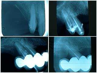
Mid- treatment radiographic x-rays series of a Smile makeover dental rehabilitation. Root canal treatment post op radiographs xrays. Note that #29 – the last tooth in the lower left x-ray – was subsequently extracted. Bottom sentence below image reads “…while waiting for the lab-processed temporaries.”
Periapical radiographs x-rays of a failing distal tooth abutment in a one year old dental rehabilitation. 1) Pre-operative x-ray . 2) This xray was taken following root canal therapy, root planing and open flap debridement. 3) This x-ray shows gutta percha placed into a periodontal gum abscess one year later. 4) This xray was taken following root planing, targeted antibiotics and tooth extraction. The roots of this extracted tooth were fused. This patient was scheduled for a three month recall; she did not return for eight months following her first three month recall visit. The periodontal abscess was readily apparent at this time. It is suggested that dental reconstruction oral rehabilitation patients are treated, not cured, and that they must be closely supervised with regular examinations and dental xrays. Patients need to be acutely aware of the need for frequent recall visits. It is possible that earlier diagnosis and intervention might have prevented the loss of this tooth.
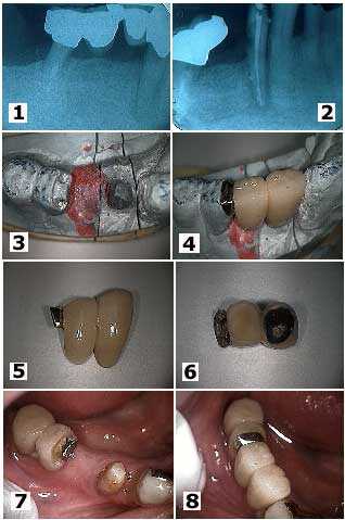
These dental x-rays and pictures show how to attach one porcelain metal teeth bridge – dental crowns – to another porcelain metal teeth bridge in the middle of a pontic. 1) Radiograph xray shows distal root dental caries under a dental bridge and a root canal infection on a lower first premolar tooth. Note that the distal molar had previously been hemisected. 2) This x-ray was taken after the bridge section was removed and root canal therapy was completed. 3) – 4) These pictures show the two and a half unit bridge fabricated and seated on the working model. 5) – 6) Two photos of the two and a half unit bridge. The distal half unit has been fabricated with a female semi-precision attachment. 7) Intra oral image. Note how the metal framework of the pontic was prepared into a “T” shape to help lock in the bridge upon cementation. 8) Photo of the final bridge.