Before and after photos on alignment of teeth with braces via leveling and aligning performed in our Braces Orthodontics office.
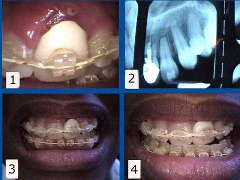
Orthodontic complications. 1) & 2) This patient presented with a failing tooth #9 under an unattractive crown. This tooth was maintained for one year while her teeth were being orthodontically aligned. The #9 tooth root was then extracted from under the crown to allow the gum and bone to heal with about eight months remaining to complete her braces. 3) & 4) This rootless crown #9 is being held in place only by the orthodontic bracket and wire. This patient preferred to keep this unattractive crown during her ortho treatment to minimize simultaneous cosmetic changes, but a cosmetic temporary crown could also have been used during this time. A dental implant will be placed about three months following the extraction. The second stage implant connection will coincide with the braces removal.
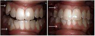
Cosmetic dental treatment of upper and lower right canine teeth. The patient declined orthodontics teeth braces. The upper and lower canines were extracted and the distal of the laterals and the mesial of the first premolars were bonded to close the space. The second photo is one week following extraction. The bonding was placed before the teeth were extracted so the patient never had to show the space between her teeth. Refer to other images in this series.
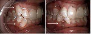
Aesthetic dentistry treatment of upper and lower right canine teeth. The patient declined orthodontics. The upper and lower canines were extracted and the distal of the laterals and the mesial of the first premolars were bonded to close the space. The second photo is one week following extraction. The bonding was placed before the teeth were extracted so the patient never had to show the space between her teeth. Refer to other images in this series.
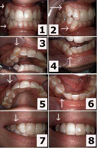
Esthetic dentistry for upper and lower right canine teeth. The patient declined dental braces. The upper and lower canines were extracted and the distal of the laterals and the mesial of the first premolars were bonded to close the space. The second photo is one week following extraction. The bonding was placed before the teeth were extracted so the patient never had to show the space between her teeth. Refer to other images in this series.
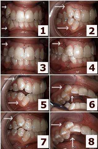
Cosmetic dentistry treatment of upper and lower right canines. Focus on the upper jaw. The patient declined orthodontics. The upper and lower canines were extracted and the distal of the laterals and the mesial of the first premolars were bonded to close the space. The second photo is one week following extraction. The bonding was placed before the teeth were extracted so the patient never had to show the space between her teeth. Refer to other images in this series.
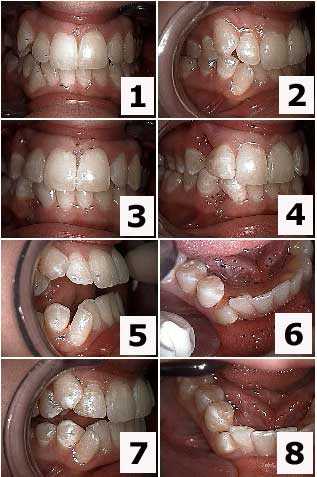
Cosmetic dental treatment of upper and lower right canine teeth. Focus on the lower jaw. The patient declined orthodontics teeth braces. The upper and lower canines were extracted and the distal of the laterals and the mesial of the first premolars were bonded to close the space. The second photo is one week following extraction. The bonding was placed before the teeth were extracted so the patient never had to show the space between her teeth. Refer to other images in this series.
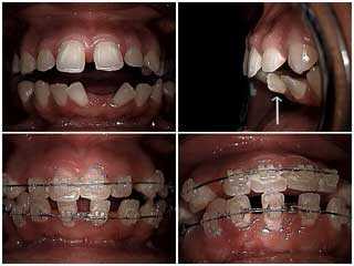
Fixed orthodontics used to retract severe labial inclination. 1) & 2) Pre-operative views. Note the upper and lower central incisors. 3) & 4) Following six months of treatment. Again, note the upper and lower central incisors. Braces move teeth through bone like moving a basketball pole through wet cement using anchorage and elastics. Note the three teeth connected with wire are being used as an anchorage unit while one tooth is being pulled by the elastic band.
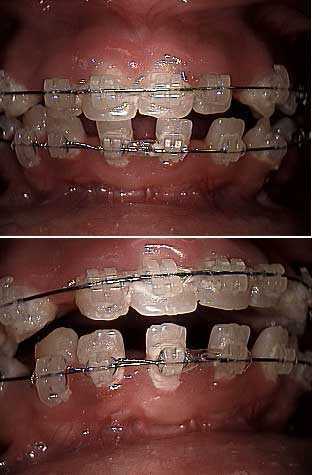
Braces move teeth through bone like moving a basketball pole through wet cement using anchorage and elastics. Note the three teeth connected with wire are being used as an anchorage unit while one tooth is being pulled by the elastic band.