Before and after photos on complications in orthodontics and problems with braces performed in our Braces Orthodontics office.
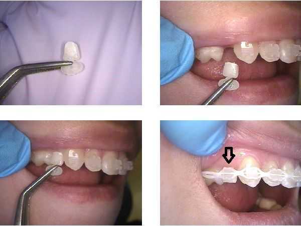
How to add a temporary crown to fixed orthodontic braces to close a gap or space between teeth. A crown form or lab-fabricated temporary crown of the appropriate shade, size and color is tried into the space to be closed. The orthodontist will then bond an orthodontic bracket to the temporary crown and attach it to the orthodontic wire using standard elastics.
How to add a temporary crown to fixed orthodontic braces to close a gap or space between teeth. This technique may be used until the space is closed with braces; the mesio-distal diameter can be reduced as the space is closed as the teeth move.
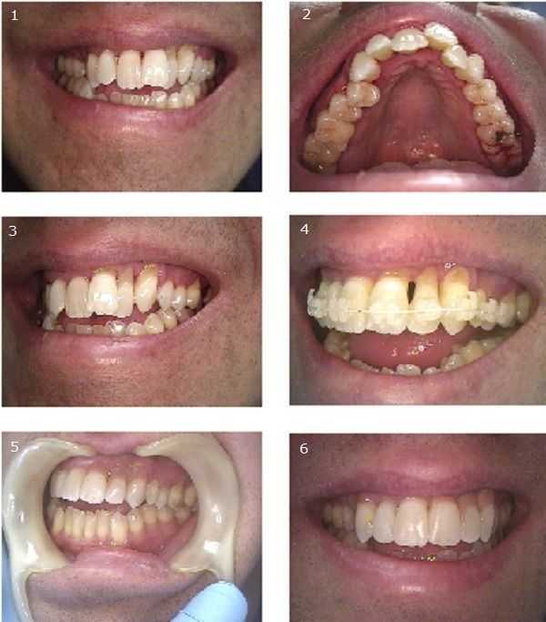
Orthodontics and Cosmetic Dentistry for a person who hates needles (who actually likes a shot, anyway?). He has received no dentistry for four years because of dental anxiety. Photos #1 & 2: The patient’s chief complaint was the crooked upper front teeth and the severe gum recession around his upper left teeth. Photos #3 & 4: The gum recession is the result of prior periodontal osseous surgery that completely disregarded esthetics. The patient did not care about his anterior open bite or crowding of the lower teeth. The patient said his anterior open bite resulted from a prior tongue thrust in childhood.
The patient consulted with our Periodontist and Orthodontist. He underwent two rounds of scaling and root planing (SRP), conservative, non-surgical gum therapy. The Periodontist suggested limiting movement of the lateral incisor #10 because it had a poor crown to root ratio; therefore the central incisor #9 was retracted more than #10 was protruded. This would also aid in reducing the anterior open bite. The patient was then cleared for limited orthodontics, teeth braces, for the upper jaw only – per patient request. The patient was put on three month recall exams with the Periodontist during orthodontic teeth movement.
Photo #4: The Orthodontist bonded the upper arch #4 – #13 (2nd premolar to 2nd premolar) with Transbond cement and placed .014 Nitinol wire. Note the cosmetic braces that use tooth-colored brackets and white wire. At patient request the orthodontic treatment plan focused on eight months of braces to align the upper arch only and not to close the open bite. After eight months the braces were removed and a bonded upper lingual retainer was placed with Transbond and GAC .017 PentaCat wire.
Photos #5 & 6: Cosmetic dentistry options for improving the appearance of the severe gum recession around the upper left teeth focused on either pink bonding at the gum line or porcelain veneers with pink gingiva (gums) at the gum line. The patient chose the pink bonding to reduce cost. This procedure did not require any shots and was completed in less than one hour.
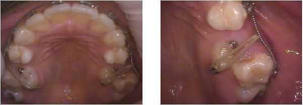
Complications in orthodontics. This patient was referred by her general dentist for a second opinion. The braces had been on her teeth for three years trying to move both palatally erupted second premolars. These teeth have not moved at all in three years but her orthodontist didn’t want to stop trying to get them to move. These teeth probably exhibit ankylosis and will never move. We agreed with the general dentist that the braces should be removed, the second premolars extracted and dental implants placed.
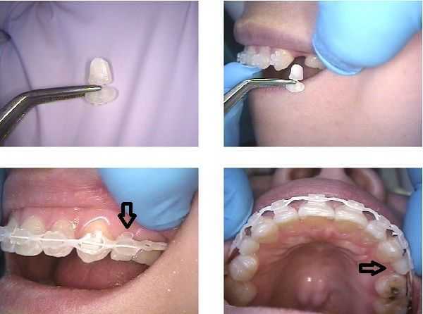
Complications in orthodontics. This orthodontic patient had a missing upper right premolar tooth – see arrow in the upper right photo. When the dental braces were placed on her teeth a non-removable false tooth pontic was added – see arrow in the lower right photo. This offered a cosmetic dental solution during teeth braces.
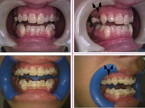
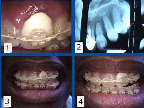
Orthodontic complications problems. 1) & 2) This patient presented with a failing tooth #9 under an unattractive dental crown. This tooth was maintained for one year during orthodontics teeth braces. The #9 tooth root was then extracted from under the dental crown to allow the gum and bone to heal with about eight months remaining to complete her teeth braces. 3) & 4) This rootless crown #9 is being held in place only by the orthodontic bracket and arch wire. This patient preferred to keep this unattractive tooth crown during her orthodontic treatment to minimize simultaneous cosmetic changes, but a cosmetic temporary tooth crown could also have been used during this time. A dental implant will be placed about three months following the tooth extraction. The second stage dental implant connection will coincide with the dental braces removal.
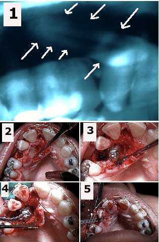
Complications in orthodontics teeth braces. Surgical exposure of a palatally impacted upper canine tooth and placement of an orthodontic bracket and elastic power chain. 1) Panoramic xray showing the horizontal tooth impaction of the upper right canine. 2) Initial oral surgery exposure of the impacted canine and extraction site after removal of an over-retained deciduous baby tooth. 3) Orthodontic bracket cemented to the canine. 4) Elastic orthodontic power chain attached to the braces bracket. 5) Emergence of power chain through the extraction site after stitching the gums. The power chain will be attached to the arch wire to pull the canine into the area of the present extraction site. The canine will assume a normal position in the arch in 18-24 months.
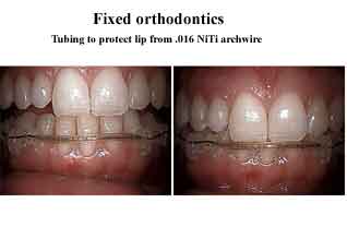
Complications in orthodontics dental braces. Tubing used to protect the lip from the teeth braces arch wire.
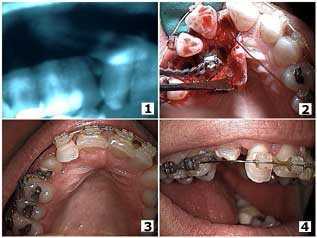
Complications in orthodontics teeth braces that requires a team approach. Top images: Orthodontic eruption of a palatally-impacted canine tooth. Bottom images: Eleven months after the surgical exposure of the canine.
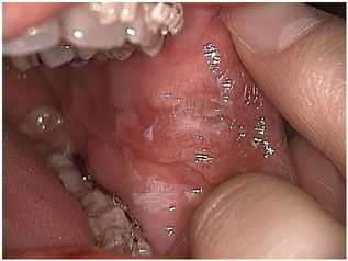
Cheek biting associated with orthodontic brackets. Complications in orthodontics teeth braces. The problem with a cheek bite is that the area will remain swollen for quite a long time and this makes it vulnerable to another cheek bite. Early intervention is important.
