Before and after photos on eruption pattern of teeth on tooth position and crowding performed in our Braces Orthodontics office.

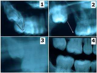
Interesting eruption pattern of a wisdom tooth. Erroneous extraction of a second molar instead of a third molar and the subsequent eruption of the third molar into the second molar position. This patient is a 27 year-old female who had recently moved to New York from Seattle. She brought prior x-rays and an interesting story involving dentistry in Seattle that I thought was worth writing about. 1) In Seattle the patient was referred to an oral surgeon for the extraction of #1 but #2 was erroneously extracted instead. 2) Radiograph after extraction of #2 showing #1 still present. 3) This new, less clear Panorex was taken a year later (still in Seattle) but the wisdom tooth #1 can be seen moving down into the second molar position on its own. 4) This radiograph was the first of the series taken in our office. The wisdom tooth #1 has almost fully erupted on its own after about two and a half years. PS – the patient was billed for and did pay for the extraction of the wrong tooth.

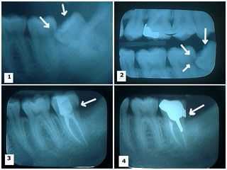
Eruption pattern of wisdom teeth and the reason for wisdom tooth extraction: damage to the adjacent second molar. 1) – 2) Two x-rays showing a lower wisdom tooth impacted on an angle and pushing into the adjacent second molar tooth causing a large tooth cavity. 3) X-ray shows the wisdom tooth was removed and the second molar following root canal therapy with the large distal tooth cavity still present. 4) Cast gold post and core and dental crown in place. Note the distal dental crown margin completely covers where the distal tooth decay was removed.

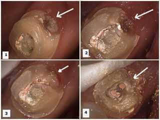
Eruption pattern of wisdom teeth – third molars – and why wisdom tooth extraction is frequently recommended: damage to the adjacent second molar tooth. These pictures show tooth preparation – drilling – of the second molar to remove the distal tooth decay after root canal therapy was performed on it and the wisdom tooth was removed. 1) – 2) Dental decay is present in the distal area of the second molar tooth. Temporary filling material is visible in the occlusal opening following root canal therapy. 3) Tooth decay removed. 4) Temporary filling material removed showing gutta percha in root canal orifices prior to cast post and core preparation.

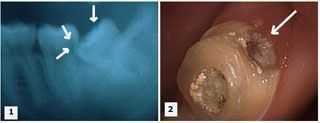
Eruption pattern of wisdom teeth – 3rd molars – and the rationale for wisdom tooth extraction: damage to the adjacent second molar tooth. 1) Xray showing a lower wisdom tooth impacted on an angle and pushing into the adjacent second molar causing a large cavity. 2) Tooth decay is present in the distal area of the second molar. Temporary filling material is visible in the occlusal opening following root canal therapy.