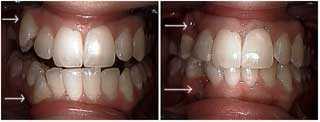
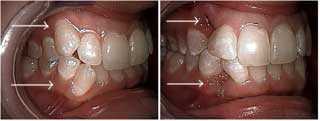
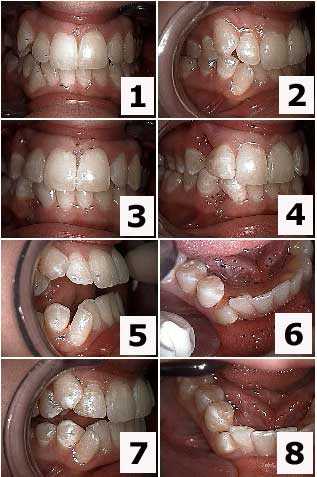
Before and after photos on malocclusion describes an unhealthy teeth bite performed in our Braces Orthodontics office.
Cosmetic dentistry treatment of an unhealthy occlusion – dental bite – of the upper and lower right canine teeth. The patient declined orthodontics teeth braces. The upper and lower canines were extracted and the distal of the laterals and the mesial of the first premolars were bonded to close the space. The second photo is one week following extraction. The teeth bonding was placed before the teeth were extracted so the patient never had to show the teeth gap space between her teeth. Refer to other images in this series.
Aesthetic dentistry treatment of malocclusion – tooth bite – of the upper and lower right canine teeth. The patient declined orthodontics teeth braces. The upper and lower canines underwent teeth extraction and the distal of the laterals and the mesial of the first premolars received dental bonding to close the teeth space. The second photo is one week following tooth extraction. The teeth bonding was placed before the teeth were extracted so the patient never had to show the tooth gap space between her teeth. Refer to other pictures in this series.
Esthetic dentistry treatment of an occlusal disharmony – a bad teeth bite – of the upper and lower right canine teeth. The patient declined orthodontics dental braces. The upper and lower canines were extracted and the distal of the lateral incisor teeth and the mesial of the first premolars received teeth bonding to close the teeth gap. The second image is one week following teeth extraction. The dental bonding was placed before the teeth were extracted so the patient never had to show the teeth space between her teeth. Refer to other photos in this series.
Cosmetic dental treatment of teeth malalignment – a bad bite – of the upper and lower right canines teeth. Focus on the upper jaw. The patient declined orthodontics dental braces. The upper and lower canines underwent teeth removal and the distal of the lateral incisors and the mesial of the first premolar teeth received tooth bonding to close the teeth gap. The second picture is one week following teeth extraction. The cosmetic bonding was placed before the teeth were extracted so the patient never had to show the teeth gap space between her teeth. Refer to other pictures in this series.
Aesthetic dental treatment of a bad teeth bite of the maxillary and mandibular right canines teeth. Focus on the lower jaw. The patient declined orthodontics teeth braces. The upper and lower canines were extracted and the distal of the laterals and the mesial of the first premolars were teeth bonded to close the space. The second photograph is one week following teeth extraction. The aesthetic bonding was placed before the teeth were extracted so the patient never had to show the teeth gaps between her teeth. Refer to other photographs in this series.
When NOT to treat a patient.
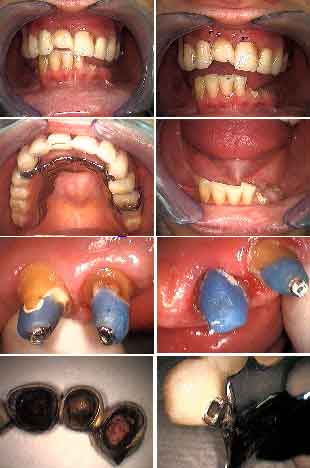
This patient presented with a loose upper fixed porcelain metal dental bridge. Her chief complaint was “to just recement it.” There is obvious tooth decay on the abutment teeth around composite post & cores. There is an obvious malocclusion – bad bite or occlusion – that was not properly addressed in this teeth bridge. This patient needs comprehensive care and was referred to a local dental school.
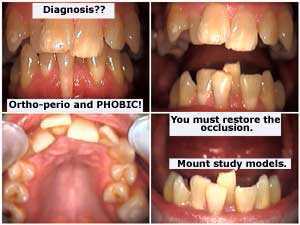
Dental Diagnosis: dental fear, orthodontics – braces, periodontics – gums and occlusion – teeth bite. You need to restore the occlusion as part of any dental treatment. Mount study models.
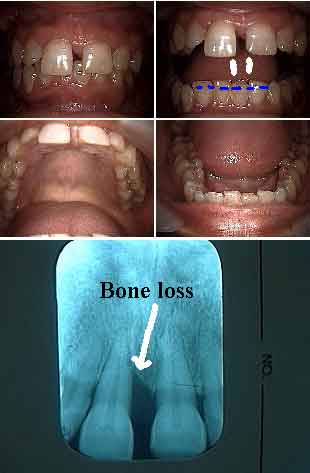
A combination of a bad bite and bad gums. Occluso-periodontal problems. This 40 year old patient’s chief complaint was her front teeth are moving and she now has a teeth gap space. Fremitus – vibration of tooth #8 upon closing the teeth was noted. Notice her deep teeth bite. Consider either lower anterior orthodontic intrusion or incisal adjustment. Next, periodontal root planing and then probably gum surgery in the upper anterior. Following gum healing consider either upper orthodontic movement – teeth braces – to bring these teeth back or dental bonding with a palatal teeth splint (after reducing #8 incisally).
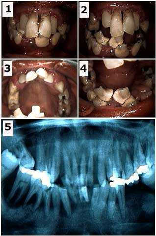
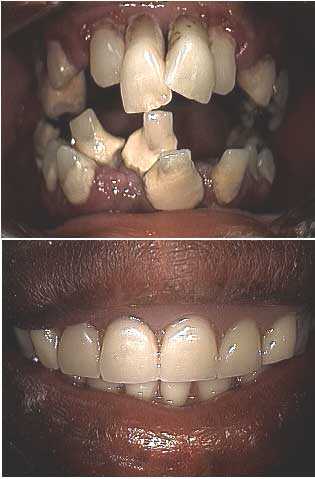
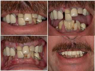
Full mouth oral reconstruction of a dental phobia patient with a severe malocclusion – bad bite or occlusion. Initial visit. It is important to determine what teeth, if any, may be saved at least temporarily. It is easier for a patient to emotionally adjust to a temporary prosthesis teeth bridge that has at least some amount of retention provided by natural teeth abutments. The teeth chosen were #6, 11, 22 and 28. The decision to fabricate a removable immediate partial denture, rather than a fixed lab-processed temporary bridge, was determined by the particular weakness of tooth #28. The patient was informed that the immediate prosthesis was to be used for the healing phase and that the four remaining abutments, particularly #28, might be subsequently extracted. Photo #2 of 4.
Before and after pictures of a full mouth oral reconstruction dental phobia patient with a severe malocclusion – bad bite.
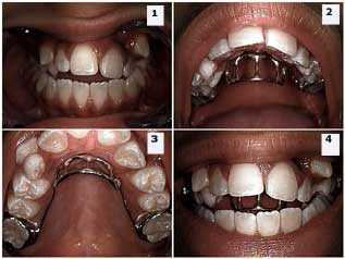
A tongue crib in place to stop a tongue-thrust habit in an adolescent male that is causing malalignment of the teeth and an unhealthy occlusion. Note the anterior open bite and the labial displacement of the upper central incisor tooth #8. The tongue crib is custom made and placed with dental cement onto the upper first molar teeth.
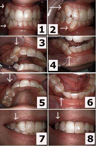
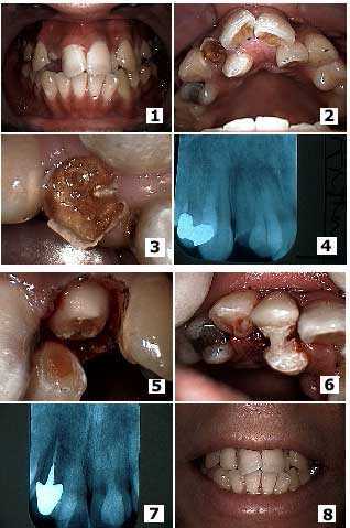
Dental malocclusion and teeth malalignment secondary to a palatally displaced supernumerary canine tooth. Treatment first focused on the adjacent broken lateral incisor tooth with severe tooth decay. Pictures 1) – 2) Initial presentation. Note the broken, decayed lateral and the palatal location of the adjacent supernumerary tooth. 3) Close up of the broken, decayed lateral and the adjacent palatally-displaced supernumerary tooth. 4) X-ray. 5) The same tooth after gum surgery and initial tooth preparation – drilling. 6) Placement of the temporary dental crown. Note that acrylic was extended from the temporary on the lateral to the supernumerary to provide initial stability of the temporary tooth crown until root canal therapy and a cast post and core was placed. 7) Radiograph of the final root canal therapy, cast post and core and porcelain crown. 8) Photos show the final before and after result.
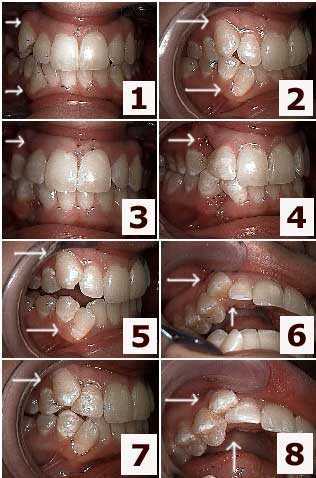
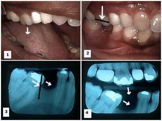
Problems with teeth occlusion – dental bite – after tooth extraction of a lower first molar tooth. This lead to supraeruption of the opposing upper molar tooth and mesial drift of the lower second molar tooth. Pictures 1) -2) The upper first molar tooth has supra-erupted because its opposing antagonist, the lower first molar tooth, has been removed. Photo 3) Mesial drift of the lower second molar tooth is seen after the extraction of the lower first molar tooth. Photo 4) Both supra-eruption of the maxillary first molar tooth and mesial drifting of the mandibular second molar tooth can be seen in this one dental x-ray.
Conclusion: tooth extraction can frequently cause significant occlusal bite problems if not actively prevented. What may have seemed like an easy and inexpensive treatment option – tooth extraction – frequently becomes a complicated and expensive repair!
Full mouth oral reconstruction of a dental phobia patient with a severe malocclusion – a bad bite or occlusion. Initial visit. It is important to determine what teeth, if any, may be saved at least temporarily.
Reconstruction performed by a Columbia University dental student in our office. The student, Jared Bowyer, is part of a unique honors program at Columbia University Dental School.
