Before and after photos on the arch length determines space for rotations performed in our Braces Orthodontics office.
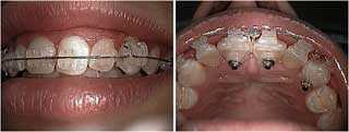
Mesio-labial rotation of tooth #’s 8, 9 & 11. Lingual buttons with elastomeric rotators.
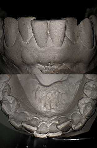
This is a frontal and occlusal view of a plaster model of the lower arch. It demonstrates teeth crowding in the lower arch. Note the malpositioned lateral incisors. These photos may also demonstrate post-orthodontic relapse associated with not wearing retainers after treatment.
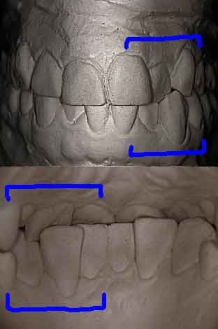
This is a frontal and lingual view of a plaster model of the same dentition. Note the anterior cross bite with the maxillary left lateral and the mandibular left canine teeth.
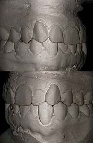
Lateral views of the same dentition. Note the anterior cross bite between the upper lateral incisor and the lower canine teeth on one side. A fixed orthodontic appliance teeth braces could be used to correct this cross bite.
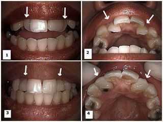
Cosmetic bonding of both upper lateral incisors to make them appear further forward. 1) Smile photo shows that the upper lateral incisors appear behind the central incisors. 2) Occlusal view of the same. 3) Post-op smile photo following bonding of the lateral incisors. 4) Post-op occlusal view shows bonding material has been added to the labial surface of the laterals to make them appear further forward.