Before and after photos on teeth space analysis for filling or closing gaps performed in our Cosmetic Dentistry office.
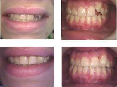
Dental space analysis for an ectopic tooth. This pretty college student had an upper left canine tooth that was crowded out of her maxillary arch. Cosmetic Dentistry with one dental bonding and one porcelain tooth veneer. Treatment time: 2 visits. The patient refused orthodontic treatment. The tooth veneer had pink porcelain around the gingival margin gums to hide its relative height.
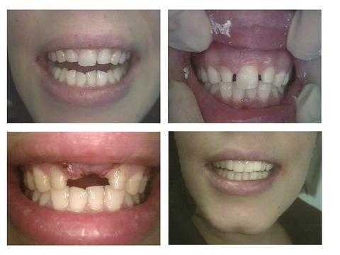
Arch length measurement and space analysis before Cosmetic Dentistry. This pretty 25 year old woman from Ireland had only one upper front tooth. She had a childhood accident and her dentist at that time extracted the broken upper right front tooth. When her other adult teeth grew in they moved forward to close the spaces and this resulted in a very odd-looking smile with only one front tooth.
After analyzing the space it was determined that if the laterals and canines were narrowed then two central incisors could be created to fit in that space. The upper lateral incisors had root canal therapy and posts placed. The laterals were then prepared for a bridge but more tooth reduction was taken from the mesial surfaces. The mesial of both canines were also reduced so that the laterals could be “distalized.” The central incisor was extracted and a bone graft was placed. A temporary lab-processed four teeth bridge is shown in this photo.This patient came from Ireland and stayed from Monday to Friday of one week. Treatment was performed in only four hours on one day with the help of an Endodontist, Oral Surgeon, Joseph Tuil Dental Lab and Cosmetic Dentist.A second lab-processed temporary teeth bridge, made by Joseph Tuil Dental Lab and reinforced with Ribbond, was placed before the patient’s departure. She will return from Ireland in three months for fabrication and insertion of the final porcelain four teeth bridge. This second visit will also occur during one week.
After analyzing the space it was determined that if the laterals and canines were narrowed then two central incisors could be created to fit in that space. The upper lateral incisors had root canal therapy and posts placed. The laterals were then prepared for a bridge but more tooth reduction was taken from the mesial surfaces. The mesial of both canines were also reduced so that the laterals could be “distalized.” The central incisor was extracted and a bone graft was placed. A temporary lab-processed four teeth bridge is shown in this photo.This patient came from Ireland and stayed from Monday to Friday of one week. Treatment was performed in only four hours on one day with the help of an Endodontist, Oral Surgeon, Joseph Tuil Dental Lab and Cosmetic Dentist.A second lab-processed temporary teeth bridge, made by Joseph Tuil Dental Lab and reinforced with Ribbond, was placed before the patient’s departure. She will return from Ireland in three months for fabrication and insertion of the final porcelain four teeth bridge. This second visit will also occur during one week.
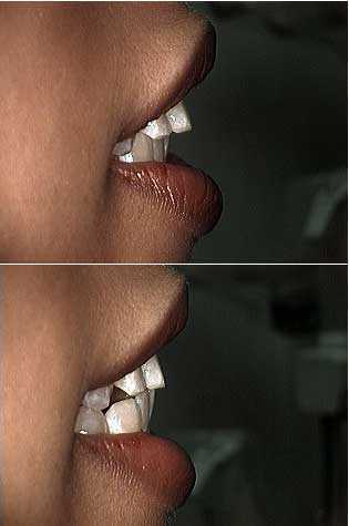
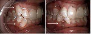
Overjet is the horizontal measure of how much the upper teeth project forward relative to the lower teeth. The first picture shows the patient’s natural teeth bite. The bite in the second picture shows how she would like to look. Orthodontics dental braces or cosmetic dentistry can achieve this. How it will occur will be partially based upon space analysis including arch length measurement and patient preferences.
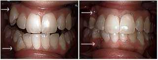
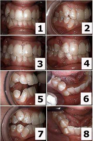
Cosmetic dentistry treatment of upper and lower ectopic right canine teeth with insufficient arch length. The patient declined teeth braces orthodontics. The upper and lower canines were extracted and the distal of the laterals and the mesial of the first premolars received dental bonding to close the space. The second picture is one week following teeth extraction. Refer to other photos in this series.
Aesthetic dentistry treatment of upper and lower ectopic teeth. The patient declined dental braces. The upper and lower canine teeth were extracted and the distal of the laterals and the mesial of the first premolars were bonded to close the gaps. The second picture is one week following teeth extraction. Refer to other photos in this series.
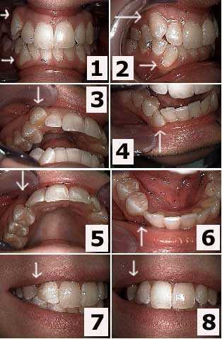
Cosmetic dental treatment of upper and lower right ectopic canines with insufficient space for proper tooth eruption. The patient declined orthodontics. How to pictures. Tooth extraction of the upper and lower canines was first performed and then the distal of the laterals and the mesial of the first premolars received teeth bonding to close the tooth gap spaces. Refer to other pictures in this series.
Cosmetic dentistry treatment of upper and lower right ectopic canines with insufficient arch length space for proper tooth eruption. How to pictures focus on the upper jaw. The patient declined dental braces. The upper and lower canines were extracted and the distal of the laterals and the mesial of the first premolars received teeth bonding to close the teeth spaces. The bonding was placed before the teeth were extracted so the patient never had to show the space between her teeth. Refer to other photos in this series.
Aesthetic dentistry treatment of upper and lower right canines with insufficient space for proper tooth eruption. How to photos focus on the lower jaw. The patient declined teeth braces. The upper and lower ectopic canines were extracted and the distal of the laterals and the mesial of the first premolars received dental bonding to close the tooth gap spaces. The bonding was placed before the teeth were extracted so the patient never had to show the space between her teeth. Refer to other pictures in this series.
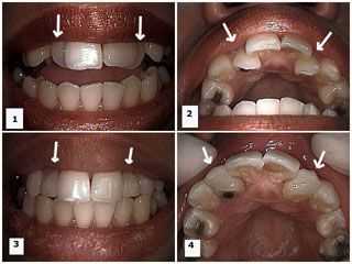
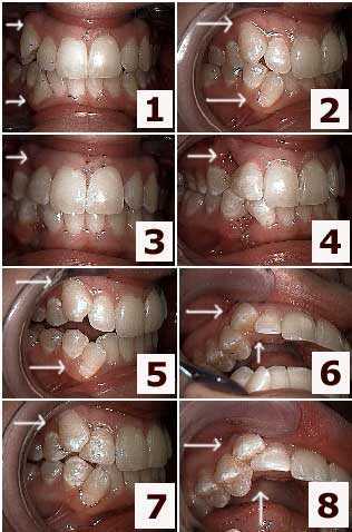
Cosmetic dental bonding of both upper lateral incisor teeth to make them appear further forward. These teeth had insufficient arch length space for proper tooth eruption. 1) Before – front photo shows the upper lateral incisors appear to be behind the central incisor teeth. 2) Occlusal view of the same. 3) Post-op front photo following teeth bonding of the lateral incisors. 4) After – occlusal photo shows bonding material has been added to the labial surface of the laterals to make them appear further forward.
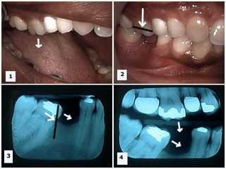
Supraeruption and mesial drift secondary to extraction of a lower first molar tooth. Pictures 1) -2) The upper first molar has supra-erupted because its opposing antagonist, the lower first molar tooth, has been removed. 3) Mesial drift of the lower second molar is seen in this x-ray secondary to the extraction of the lower first molar. 4) Both supraeruption of the upper first molar and mesial drifting of the lower second molar can be seen in this one xray following the removal of the lower first molar. Conclusion: tooth extraction can frequently cause significant changes in arch length and teeth spacing if not actively prevented.