Our tooth extraction and dental implants team includes 16 cosmetic dentists and specialists, including two oral surgeons, in one high-end office. In particular our tooth extraction and dental implants treatment is quick and comfortable. In addition our
Our tooth extraction and dental implants patients also have access to top medical care
Moreover our patients have access to our affiliated team of 12 medical doctors in our building. This is because many patients who need a tooth extraction also tend to neglect their overall medical health. Above all we hope to improve your overall health comprehensively.
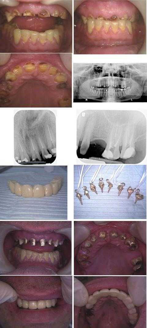
Cosmetic dental and tooth extraction
Total treatment time was about nine hours in two visits during one business week: Monday (four hours) and Friday (five hours). This patient hid his dental condition for over a decade by never smiling.
In the first row of photos one can see that the bite was over-closed. The patient had a prior history of an eating disorder. In particular this patient also had a current tooth grinding habit.
A panoramic x-ray is seen in the second row of photos. An Oral Surgery consultation was performed prior to treatment. This was to evaluate opening the bite.
The third row shows x-rays taken after the Endodontist completed eight root canals. Especially this was accomplished on the first day of treatment. The treated teeth include tooth #’s: 6 – 11 and 14, 15.
The fourth row shows the lab-processed 11 unit (11 tooth) temporary bridge. It also shows the cast gold post and cores (with Kaitlyn loops) for those teeth that received root canal treatment.
Next the fifth row shows the cast gold post and cores after cementation.
Then the sixth row shows the lab-processed temporary bridge in place. By the way the oral surgeon extracted tooth #’s: 3 – 5 and 12. crown lengthening gum surgery was performed on tooth #’s: 6 – 11. In addition a distal wedge gum surgery was performed on #15.
Lastly the patient will have a final porcelain-metal bridge made after the gums heal. Dental implants may also be placed in the upper right posterior. However saving these teeth gave the patient a choice to use or not use dental implants in his dental reconstruction. Above all a bite plate is also necessary to try to mitigate the force of tooth grinding. Furthermore a referral for pharmacological management of anxiety is also worthwhile.
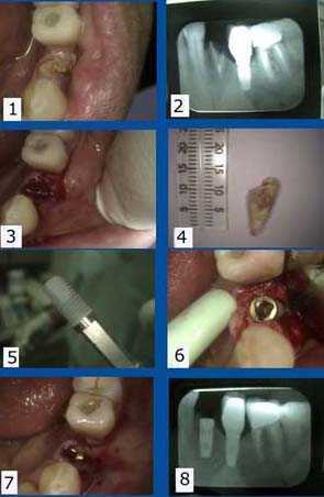
Dental implants surgery after tooth extraction
How to Pictures. The lower left second premolar position already has a dental implant as well. 1) – 2). Pre-op pictures show the fractured tooth clinically and in the x-ray. 3) Next the tooth socket is exposed and ready for placement of the dental implant. 4) Then the tooth was extracted.
Single tooth dental implant placement after tooth extraction of a fractured but not infected lower left first premolar
5) A Replace titanium threaded dental implant ready for insertion.
6) The dental implant was inserted into the mandible using a slow speed handpiece.
7) The implant was covered by the surrounding gum tissue. It was sutured using 3-0 chromic gut dissolvable sutures. 8) Lastly the post-op x-ray is shown.
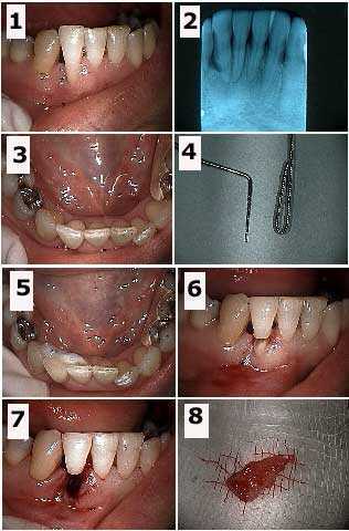
Tooth extraction of the root of a hopeless tooth #26 and splinting the coronal portion to the adjacent teeth
1) – 3) Initial images. 4) The wire used for the teeth splint is placed next to a periodontal probe. The wire splint is doubled to prevent the tooth from rotating around it after placement. 5) Then the splint is bonded to the lingual surfaces of teeth #’s 25 and 27. 6) Initial preparation – drilling – through the mid-length of the tooth. 7) Next this picture was taken following extraction of the tooth root. 8) Finally the extracted tooth root is shown.
In summary this patient has already been through two rounds of scaling and root planing. She will then have an apically positioned flap periodontal gum surgery and a bone graft after healing of this tooth extraction site. Subsequently a Maryland bridge was placed. In particular this offered the patient an alternative to dental implants following tooth extraction.
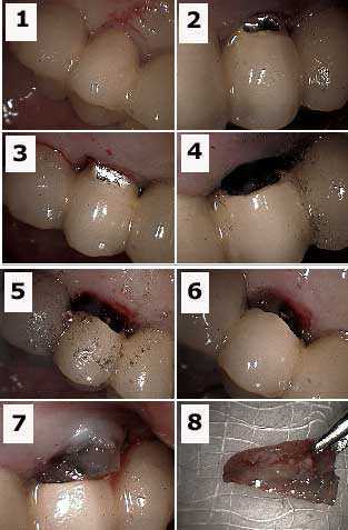
Technique for tooth extraction or root resection from under a porcelain fused to metal dental bridge
1) Labial photo of an abutment tooth with an endo-perio (endodontic – periodontal) lesion or infection. 2) & 3) Initial preparation into porcelain with a diamond bur. 4) Labial photo showing preparation. The metal portion of the dental bridge is prepared, i.e. drilled with a steel bur.
5) The palatal photo shows the tooth preparation. 6) The labial photo shows the tooth root under the porcelain. Gutta percha is visible in the tooth root. 7) Then the tooth root is extracted. Moreover the occlusal height of this root must first be reduced to allow it to be removed. 8) The extracted tooth root.
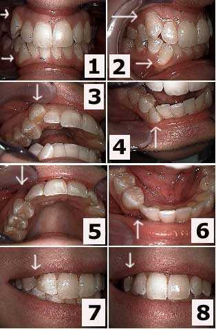
Cosmetic dentistry treatment of upper and lower right canines by dental extraction
The patient declined orthodontics tooth braces. The upper and lower canine teeth were extracted and the distal of the laterals and the mesial of the first premolars received teeth bonding to close the gap. The second photo is one week following tooth extraction.
In this case the dental bonding was placed before the teeth were extracted so the patient never displayed a space between her teeth. Refer to other pictures in this series.
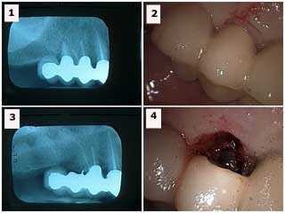
Tooth root extraction resection from under a porcelain fused to metal teeth bridge
1) The X-ray radiograph shows the distal tooth abutment with a combined root canal and gum infection (endo-perio lesion). 2) The labial photo of the same tooth is seen. 3) Radiograph following tooth root resection. The metal chad in the area of the extracted abutment was later removed. 4) The labial picture following teeth extraction.
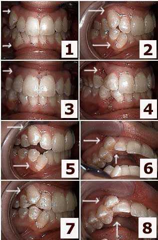
Esthetic dentistry with teeth extraction for a patient who declined dental braces orthodontics
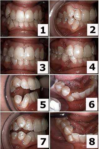
Aesthetic dentistry for a patient who refused teeth braces orthodontics and instead chose teeth extraction
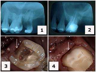
Extraction of an upper molar with severe dental caries – tooth decay cavity
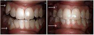
Before and after pictures of cosmetic dental treatment and teeth extraction
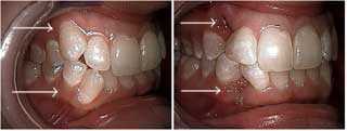
Before and after pictures showing cosmetic dentistry treatment for a patient who declined dental braces
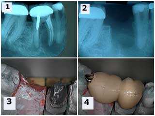
How to build an occlusal rest in a dental crown and avoid removing a dental crown after tooth extraction
This two tooth dental bridge has an occlusal rest seat seated onto the rest preparation in the mesial of the second molar dental crown. This eliminates the need for removing the second molar dental crown. The premolar tooth did receive root canal therapy following healing of the tooth extraction site. This avoided the patient needing dental implants after tooth extraction.
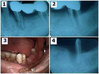
Extraction of a mandibular premolar tooth and tooth root resection of a molar with furcation involvement following an accident
1) The initial x-ray shows the three teeth bridge and severe damage to the second premolar tooth. 2) The second molar tooth shows a furcation involvement – advanced gum disease – and thickening of the periodontal ligament space around the mesial tooth root. The second premolar tooth and the mesial root of the second molar tooth were extracted. 3) This intra oral photo was taken after healing of the extraction site. The patient was informed regarding the long span and declined dental implants. He wanted a new fixed dental bridge anyway. 4) Radiographic healing of the distal root of the second molar after six weeks. The lesson here is the value of salvaging individual tooth roots of molars. Again this allowed the patient to avoid dental implants after tooth extraction.
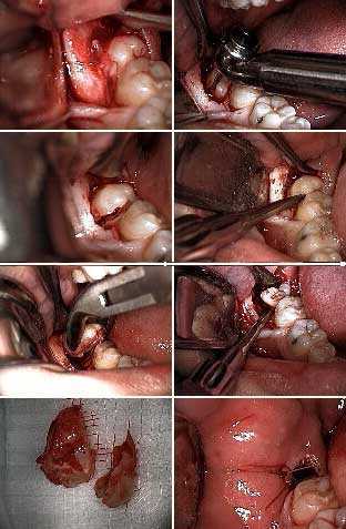
How to pictures show wisdom tooth extraction – a lower right partial bony impacted wisdom tooth
1) This photo shows soft tissue reflection. 2) & 3) The molar tooth is sectioned. 4) Then an elevator is used to split the molar.
5) & 6) The tooth roots are split using a lower forceps and an elevator. 7) Next the extracted tooth is shown in two sections. 8) The surgical site is then closed with stitches.
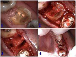
Surgical tooth extraction of a hopeless upper first molar
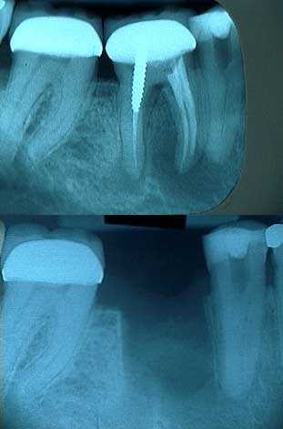
Rationale for tooth extraction
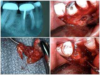
Pictures show tooth extraction of a mandibular molar tooth #30 with severe periapical pathology – tooth abscess infection around the tooth roots
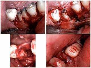
Extraction of tooth #30 due to severe periapical – endodontic – oral pathology
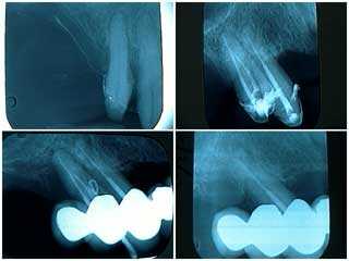
X-rays of a failing distal tooth abutment in a one year old full mouth reconstruction and need for tooth extraction
1) Pre operative X-ray of the upper right jaw. 2) In this case root canal, periodontal scaling and open debridement were initially performed. 3) Subsequently the patient returned one year later with a fistula or gum boil around one tooth. Therefore gutta percha was placed into the periodontal gum abscess to track its origin. 4) Next scaling was performed along with targeted antibiotics. Then tooth extraction was performed. The roots of this extracted tooth were fused.
Ordinarily this patient should have remained on a three month recall. However she did not return for eight months following her first three month recall visit. As a result the periodontal gum abscess occurred. It is suggested that oral reconstruction patients are treated, not cured. Furthermore they must be closely supervised. Patients need to be acutely aware of the need for frequent recall visits. In particular it is possible that earlier diagnosis and intervention might have prevented the loss of this tooth.
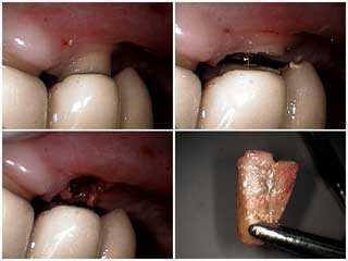
How to pictures method for extracting a failing distal tooth abutment from under a porcelain fused to metal dental bridge reconstruction
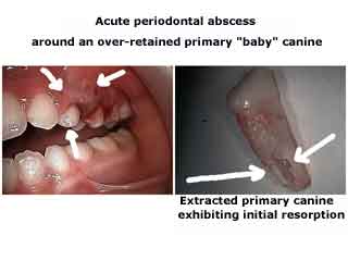
Baby tooth extraction. Primary tooth extraction means the same thing
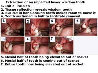
Wisdom tooth extraction
How to pictures show surgical extraction of an impacted lower wisdom tooth. Technique.
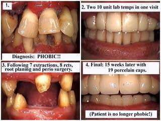
Full Mouth Oral Reconstruction – Smile Makeover and tooth extraction
In particular the diagnosis was dental anxiety dental fear.
In this case treatment consisted of: 7 teeth extractions, 8 root canals and composite cores. Next full mouth scaling and then gum periodontal surgery were performed. Finally the last picture shows 19 teeth of dental bridgework over 8 teeth abutments. In particular all dental treatment was completed in 15 weeks.
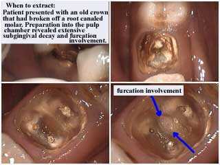
When to extract a tooth – when is tooth extraction needed
This patient presented with an old dental crown that had broken off a molar. It had previously received root canal therapy. Tooth preparation i.e. drilling into the pulp chamber revealed extensive subgingival tooth decay cavity or dental caries. Furthermore this cavity extended through the bottom of the pulpal floor resulting in furcation involvement. In summary tooth extraction is indicated. Then a dental implant should be placed after up to four months of healing.
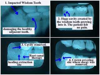
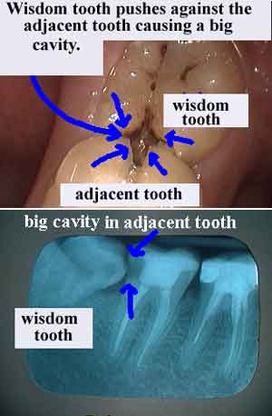
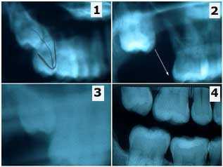
One reason wisdom teeth extraction is often needed
In these xrays an impacted wisdom tooth damages the healthy adjacent second molar tooth. A large tooth cavity was created by the wisdom tooth growing into it. The patient did not feel tooth pain.
Incidentally treatment included root canal and crown. The crown covers the site where tooth decay was removed.
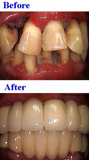
Before and after pictures of Full Mouth Oral Reconstruction involving many teeth extractions
In this case treatment included 7 extractions, 8 root canals and composite cores. Next full mouth scaling and root planing and then periodontal surgery. Lastly 19 teeth of dental bridgework was made over 8 teeth abutments.
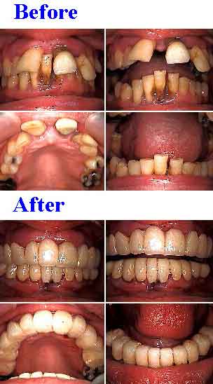
Full mouth oral rehabilitation – or smile makeover involving many teeth extractions on a dental anxiety dental fear patient
So then 20 temporary dental crowns, 14 teeth receiving root canal and 6 teeth extractions were all completed in one visit.
In summary enough natural teeth were retained for fixed porcelain caps in both jaws. Therefore dental implants were not needed following tooth extraction.
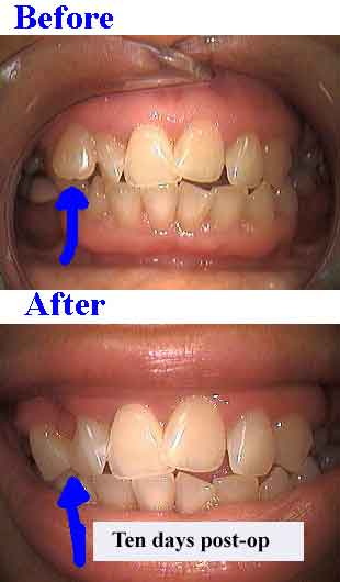
Supernumerary tooth extraction and cosmetic dentistry with dental bonding
In this case total treatment time was one visit!
The supernumerary tooth was first prepared to open the interproximal area between the remaining teeth. Then teeth bonding was placed interproximally on both teeth as if the supernumerary was already gone. This gives a clean field. Lastly the supernumerary tooth was extracted.
Wisdom teeth extraction
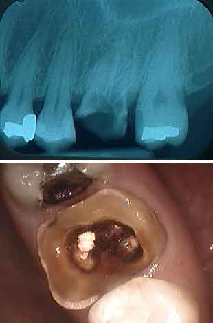
Extract this tooth
In the xray the tooth decay can be seen extending well below the level of the bone around the tooth. This tooth decay i.e. dental caries can be seen in the photo.
Tooth extraction and placement of a dental implant is indicated.
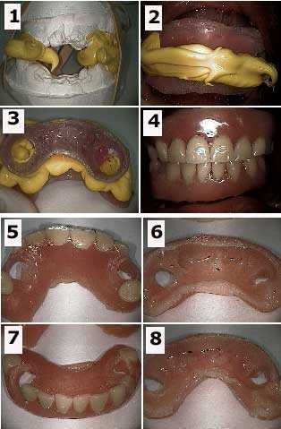
Multiple teeth extractions with dental sedation during the beginning phase of a smile makeover
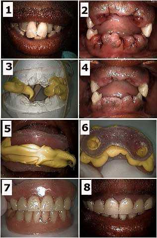
Multiple Teeth Extractions in a cosmetic dental sedation patient
It is easier for a patient to emotionally adjust to a temporary prosthesis that has at least some amount of retention provided by natural tooth abutments. The teeth chosen were #6, 11, 22 and 28.
Furthermore the decision to fabricate a removable immediate partial denture, rather than a fixed lab-processed temporary bridge, was determined by the particular weakness of tooth #28.
1) Initial. 2) Following teeth extractions of all but the four remaining tooth abutments. 3) Working models and bite made right after the dental extractions. This will serve for the fabrication of the immediate denture. 4) Three days following the teeth extractions. 5) Intra oral photo of bite rims and bite material. 6) Extra oral photo of bite rims and bite material. The patient did not want any wire clasps to be used to retain the prosthesis. 7) Extra oral image of the immediate dentures. 8) Intra oral image of the immediate dentures.
A soft reline material was used on the gingival side of these dentures. This soft reline material was allowed to flow into the circular openings (see photo #6) in the acrylic denture as it was seated. This soft reline material provided retention instead of using denture clasps. This took two visits over three days.
In summary enough natural teeth were retained in both jaws to provide adequate retention of the removable partial dentures. Therefore dental implants were not needed following tooth extraction.Photo #3 of 4.
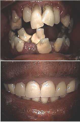
Cosmetic dental before and after pictures of a dental phobia patient that involved teeth extractions
Impacted wisdom tooth extraction
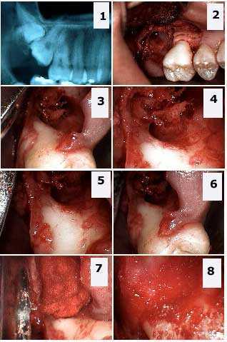
Extraction of two impacted teeth: the upper right third molar i.e. the wisdom tooth and the second molar. 1) An X-ray shows the double teeth impaction. 2) – 6) For example note different views of the large osseous defect in bone and the significant exposure of the distal furcation of the first molar. 7) – 8) Then packing the defect with freeze-dried bone and gelfoam.
Tooth extraction complication – buccal dehiscence
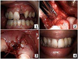
Surgical closure of a buccal dehiscence on an upper lateral incisor. 1) The initial presentation with the lateral incisor root exposed. In particular note tooth decay up to the tooth root apex. 2) Surgical exposure of the site after extraction of the tooth root. A full thickness mucoperiosteal flap with bucco-lingual vertical releasing incisions was used to obtain primary closure. After the tooth socket was curetted then freeze-dried cortical bone chips were placed with Biomend membrane. 3) Primary closure was obtained with 3-0 Vicryl sutures. 4) Finally the result after fabrication of the porcelain-metal fixed bridge. The patient chose a fixed bridge rather than a dental implant because it could be completed in much less time.
Baby tooth extraction aka primary tooth extraction then dental implant surgery
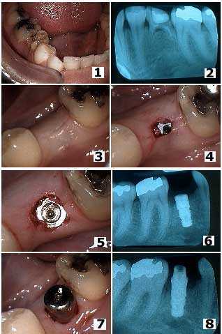
Removal of a retained primary molar tooth and placement of a dental implant. 1) Intraoral photo of the retained primary molar in the second premolar position. 2) X-ray of this tooth. Note that these retained primary teeth can remain in the mouth many years. This tooth was finally getting loose in this 37 year-old female. 3) Primary closure after removal of the tooth and dental implant placement.
Above all, the dental implant was placed at the same time as the extraction because the extracted tooth was not infected and its removal did not leave a void in bone. 4) Initial location of the first stage dental implant four months later. 5) Occlusal view after removal of the healing screw at four months. 6) Radiograph at four months. 7) The dental implant healing collar in place. 8) X-ray of the dental implant with healing collar in place.
Adults with healthy baby teeth i.e. primary teeth should not consider tooth extraction and then placement of dental implants. It is possible for healthy baby teeth to function throughout adult life.
Tooth extraction mistake – the wrong tooth was extracted
Mistaken tooth extraction of a second molar tooth instead of an impacted wisdom tooth – third molar – and the subsequent eruption of the third molar into the second molar position. This patient is a 27 year-old female who had recently moved to New York from Seattle. Accordingly she brought prior x-rays and an interesting story involving dentistry in Seattle that I thought was worth relating.
1) In Seattle the patient was referred to an oral surgeon for the tooth extraction of #1 but #2 was mistakenly extracted instead. 2) Subsequently a radiograph was taken after extraction of #2. It shows #1 is still present. 3) This new, less clear Panorex was taken a year later (still in Seattle) but the wisdom tooth #1 can be seen moving down into the second molar position on its own. 4) This x-ray was the first of the series taken in our office. The wisdom tooth #1 has almost fully erupted on its own after about two and a half years. PS – the patient was billed for and did pay for the extraction of the wrong tooth.
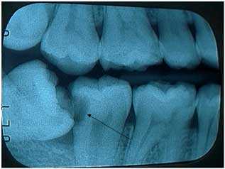
Remove impacted wisdom teeth – third molar teeth – before they damage adjacent teeth
The lower wisdom tooth is on an angle and leaning against the second molar. This has caused big tooth decay – dental caries – in the second molar. Incidentally that cavity will require root canal therapy and a dental crown to repair. In the meantime the patient did not feel tooth pain.
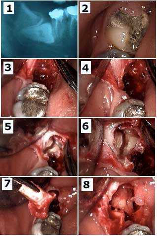
Teeth extractions – removal of a horizontally impacted lower wisdom tooth and a second molar with a lot of tooth decay
The patient was in a lot of dental pain. 1) Pre-op x-ray. 2) Tooth #31 had a temporary dental filling in it. 3) – 4) After removal of the dental crown of #32 showing the residual empty tooth socket. 5) – 6) Next a minimal gum flap is reflected to show exposure of the tooth roots of #32 with sectioning. 7) Then removal of first tooth root. 8) Thereby the second tooth root is brought forward in the tooth socket and ready for removal.
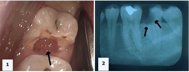
A hopeless tooth that should be extracted
1) In this case the picture of tooth #18 shows gum tissue growing into the gap previously occupied by tooth structure. 2) X-ray showing extensive tooth decay extending below the level of the gum and jawbone. Thus placement of a dental implant should following this tooth extraction in several months.
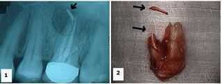
Root canal failure results in tooth extraction
1) X-ray shows an upper first molar with gutta percha – root canal filling material – extruded into a large radiolucent area. 2) Palatal photo of the extracted tooth shows the extent of the gutta percha extrusion. Namely the extra fragment shown is also gutta percha.
This tooth extraction site should be left to heal for at least four months before placement of a dental implant. This allows time for bone to fill in the tremendous bony defect that resulted from this chronic infection.
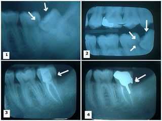
The reason for wisdom teeth extraction
These x-rays explain the reason for wisdom tooth – third 3rd molar extraction. For instance damage to the adjacent second molar tooth is shown. 1) – 2) Two xrays show a lower wisdom tooth impacted on an angle and pushing into the adjacent second molar causing a large tooth cavity. 3) Next the X-ray shows the wisdom tooth was removed and the second molar following root canal therapy. In this case a large distal tooth cavity is still present. 4) Lastly the cast gold post and core and crown was placed. In particular note the distal crown margin completely covers where the distal tooth cavity was removed.
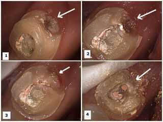
Why wisdom teeth extraction is needed
In this case these pictures explain why wisdom teeth – third 3rd molars – should be extracted. There is damage to the adjacent second molar tooth. These photos show preparation of the second molar to remove the distal dental caries after root canal was performed on it and the wisdom tooth was removed. 1) – 2) Dental caries is present in the distal area of the second molar tooth. Temporary dental filling material is visible in the occlusal opening following root canal therapy. 3) Then the tooth decay is removed. 4) Lastly the temporary filling material is removed. It shows gutta percha in root canal orifices prior to cast post and core preparation.
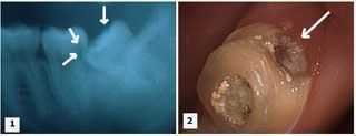
Wisdom teeth impaction extraction
In conclusion Dr. Jeffrey Dorfman created all of the dentistry shown on this 4,400 page website. We offer expert tooth extraction and dental implant placement. In brief we offer intelligent & honest diagnosis and better results for our patients. Visit us when you want it done right the first time; you will save money by initially spending more. Therefore please call The Center for Special Dentistry®.