Before and after photos on abutment teeth and dental bridges and crowns performed in our General Dentistry office.

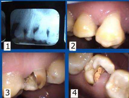
Tooth #13 has been left exposed for over a year since the root canal temporary filling fell out. 1. Current x-ray. 2. Buccal view. 3. Palatal view. 4. Occlusal view. Treatment options: Extraction and then tooth replacement or retreat the root canal, post and core, crown lengthening gum surgery and crown.

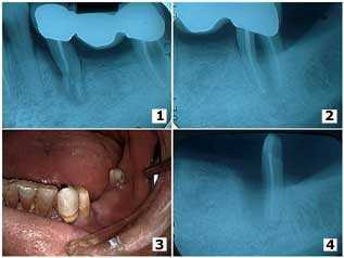
Trauma from a fall in an elderly male resulted in tooth root fracture of the mesial abutment of a three-unit dental bridge and a furcation involvement in the distal molar abutment. 1) – 2) The pre-operative x-rays. 3) The second premolar was extracted and the first premolar was used as the mesial abutment of a new four-unit bridge. 4) In this picture the second molar was hemisected because of deep probing depths around the mesial root and the mesial root was extracted. The distal root was used as the distal abutment of the new bridge. Bone fill can be seen around the mesial surface of the distal root; a cribriform plate and periodontal ligament space can be seen developing.