Photos on fix broken porcelain caps created in our General Dentistry office.

How to repair a porcelain crown. People frequently chip the porcelain on their dental crowns. Many want to repair, rather than replace, the crown especially if the crown is attached to many other crowns. Porcelain cannot be attached to the area where the metal is exposed so the only option is to use bonding material. The problem is that bonding will not chemically adhere to the exposed metal. Therefore one must prepare (i.e. drill) more of the porcelain away around the exposed metal (seen on the left) and bond an appropriate color to the entire area. The long term prognosis of this type of repair depends upon many factors.
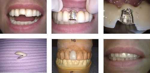
How to photos show repair of broken porcelain that is part of a large fixed porcelain metal bridge – caps. This technique can help prevent the need to replace an entire bridge. Fabricate a 3/4 crown to fit over the metal frame of the 12 teeth fixed porcelain metal dental bridge. Use dental cement to attach it.
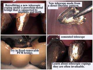
Fabricating a telescopic coping under a porcelain-metal dental bridge that is supported by implants and teeth.
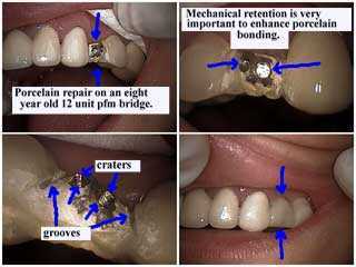
Porcelain repair on an eight year old 12-tooth PFM dental bridge. Note the grooves and craters. Mechanical retention is very important to enhance porcelain bonding.
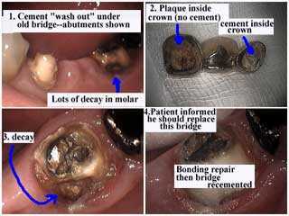
Cement leakage – washout – under old dental bridge. Rampant tooth decay may be present on the abutment teeth upon removal of a loose bridge. The words, crown and cap, mean the same thing; a dental crown and tooth crown also mean the same thing. A fixed dental bridge, prosthesis or bridgework, typically means more than one crown – and that they are connected together as one piece. The word fixed typically means permanently cemented and not removable by the patient. Some patients incorrectly refer to their removable denture as a bridge.
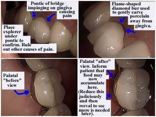
This dental bridge pontic was impinging – pushing – on the gum causing pain. Blanching of the gingiva, the gum turning white, was noted in this area. How to reduce a pontic that is pushing on the gums.
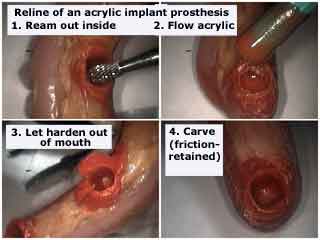
These pictures show how to reline an acrylic dental implant prosthesis.
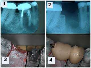
Two-unit teeth bridge with an occlusal rest seats onto the rest preparation in the mesial of the second molar crown. This eliminates the need for removing the second molar dental crown. The premolar did receive root canal therapy following healing of the extraction site.
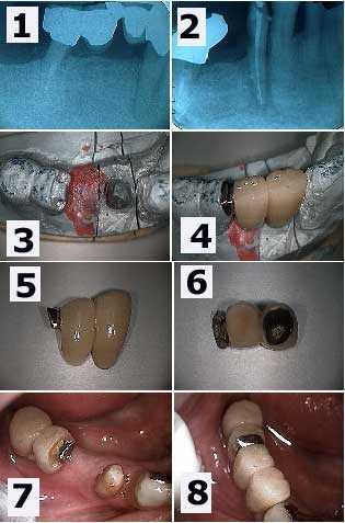
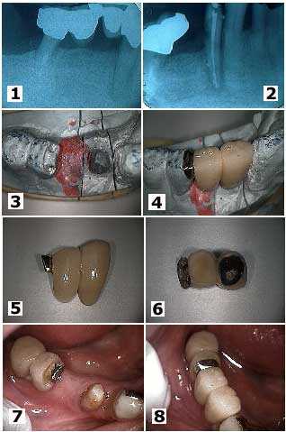
Cervical decay and root canal infection around a mandibular premolar. 1) A large radiolucency is seen in the x-ray. The distal abutment is actually the distal root of a first molar following hemisection. 2) Following root canal therapy. The cervical decay can now be seen more easily. 3) The working model showing the preparation of the mesial part of the distal two tooth dental bridge. This preparation will allow rigid connection to the new anterior bridge being made. 4) The new bridge section in place on the working model. 5) & 6) Two pictures of the new bridge showing the distal attachment. 7) An intra oral photo, which mirrors the working model view seen in 3). 8) The final result. Porcelain could have been added to the gold occlusal rest upon patient request.
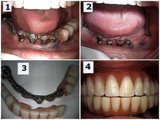
Acrylic failure in an acrylic fused to metal full arch dental implant prosthesis, dental bridge. 1) The acrylic separated from the metal of this eight-year-old implant prosthesis. 2) The dental implant abutments are shown following removal of the prosthesis. 3) The prosthesis showing the separation. 4) Following fabrication of new acrylic to the frame. The ability to remove, repair and replace a prosthesis is a good thing.
How to attach a dental bridge to another dental bridge in the middle of a pontic. Pictures. 1) This elderly patient developed distal root decay under a bridge and a root canal infection on an abutment premolar. Note that the distal molar had previously been hemisected. 2) Following dental bridge section removal and endodontics. 3) – 4) Two and a half unit bridge fabricated and seated on the working model. 5) – 6) Two photos of the two and a half unit bridge. The distal half unit has been made with a female semi-precision attachment. 7) The metal framework of the pontic was prepared into a “T” shape to help lock in the bridge upon cementation. 8) The final porcelain metal dental bridge.
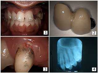
Dental bridge repair. This patient could not afford replacement of the bridge. 1) – 2) Patient had a two tooth bridge on an upper canine abutment, lateral incisor pontic and palatal wing attached to the distal of the central. The patient presented with the bridge out. 3) Initially it seemed that there was external resorption on the facial surface of the canine near the gingival margin. 4) Initial x-ray. Photo #1 of 3.
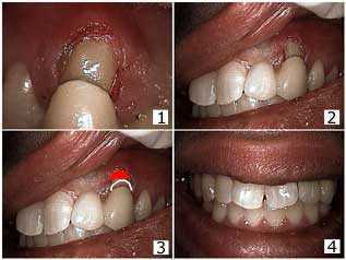
Dental Bridge repair. The patient could not afford replacement of the bridge. 1) Fitting the dental bridge over the bonded repair. 2) The bridge was cemented with resin. The mesial wing was also bonded to the distal of the lateral. 3) The gingivectomy was chosen over a periodontal gum flap surgery because the patient had a low lip line and so the development of a pseudopocket with a flap was avoided. The red indicates where the gum line could have been. 4) The final result. The patient is informed of the need for bridge replacement when finances allow. Photo #3 of 3.