Photos on Pediatric dentists treat primary or baby teeth created in our General Dentistry office.
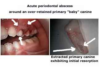
Acute periodontal gum abscess infection around a deciduous baby canine tooth that needed tooth extraction by a Pediatric Dentist. The extracted baby tooth is seen in the next photo.
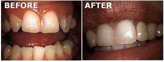
Before and after pictures show a porcelain dental crown – cap – placed on an upper peg lateral incisor tooth #10. Root canal therapy was also performed. Peg laterals are not usual.
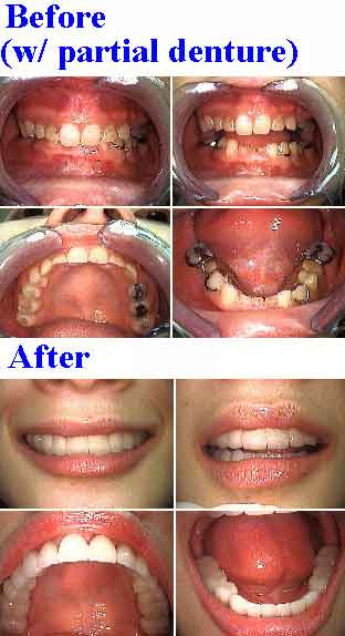
Full Mouth Dental Reconstruction – oral rehabilitation – for a 30 year old female who had many missing adult teeth, retained baby teeth – partial anodontia – and a closed bite occlusion. Before and after pictures taken three weeks apart. A Pediatric Dentist was unable to help this patient in childhood resulting in the need for a smile makeover before attending law school.
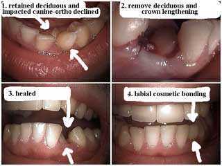
How to cosmetic dentistry pictures show treatment for a retained baby tooth and an impacted adult canine tooth. After tooth extraction and healing of the deciduous baby tooth the labial surface of the impacted adult canine tooth received cosmetic dental bonding to close the remaining tooth gap space. The patient refused pediatric dentist advice as a child and refused orthodontics as an adult.
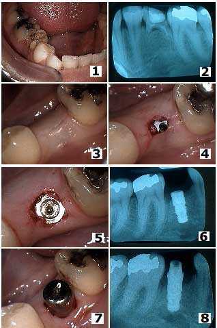
Tooth extraction of a retained deciduous primary baby tooth and placement of a dental implant in a 37 year old female. How to pictures. 1) Intra oral photo of the retained baby tooth in the second premolar position. 2) Radiograph x-ray of this tooth. Retained primary baby teeth can remain in the mouth many years; this primary tooth was finally getting loose in this 37 year old! 3) Primary closure after tooth removal and dental implant placement. The dental implant was placed by the Oral Surgeon at the same time as the tooth extraction because the extracted tooth was not infected and its removal did not leave a void in the jaw bone. 4) This photo shows the initial location of the first stage dental implant four months later. 5) Occlusal photo after removal of the dental implant healing screw at four months. 6) Radiograph x-ray at four months. 7) The dental implant healing collar in place. 8) Radiograph xray of the dental implant with healing collar in place.