Photos on dental radiographs X-rays created in our General Dentistry office.
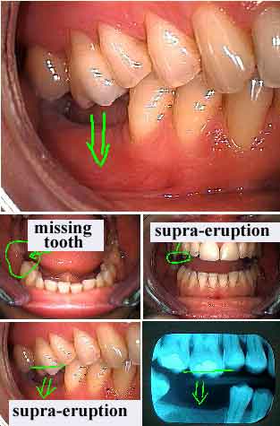
Dental supra-eruption occurs when a tooth continues to grow out of the gum if the opposing tooth in the opposite jaw is missing. Treatment involves placing a tooth in the missing space and adjusting the height of the supra-erupted tooth. The digital dental x-ray shown here among the pictures clearly shows both tooth supraeruption and mesial drift of the lower back molar tooth.
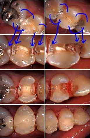
Dental x-rays are valuable in diagnosing tooth decay in teeth which appear to be clinically – visibly – healthy. Digital dental x-ray diagnosis is more difficult than with film. This is true for all types of dental radiographs: periapical, bitewing or panoramic.
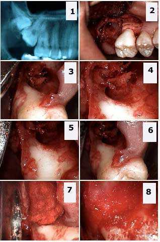
This digital dental panoramic x-ray shows two maxillary molar teeth impactions. The pictures show the extraction of both the wisdom tooth and second molar tooth. 1) Dental radiograph shows the double tooth impaction. 2) – 6) Different photos show the large osseous defect in the jaw bone and the significant exposure of the distal furcation of the first molar tooth. 7) – 8) Packing the defect with freeze-dried bone and gelfoam.
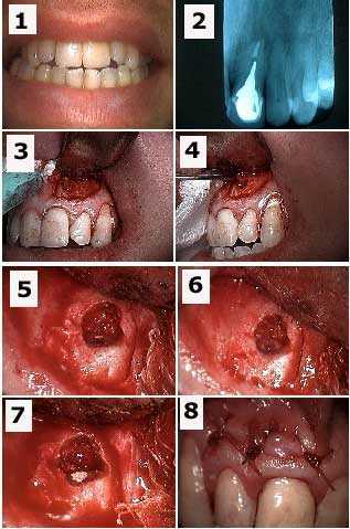
Dental x-ray taken before oral surgery. This patient had a draining fistula gum boil. Gutta percha was placed in the fistula and a dental radiograph was taken – not shown here – to identify the origin of the tooth infection. Pictures of the apicoectomy are shown here. Gutta percha can be gently placed into a draining fistula without local anesthesia.
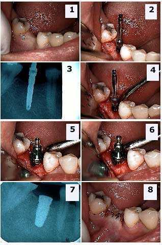
Dental implant oral surgery for the first stage tooth implant in the lower first molar position. The abutment teeth have minimal dental restorations. 1) The pre-operative photo. The clinical exam revealed sufficient width and depth of bone for the placement of the dental implant. 2) An osteotomy was made with a Sterioss bur with internal irrigation. 3) A dental radiograph x-ray was taken to ensure enough room between the apex of the implant and the inferior alveolar nerve. 4) The Sterioss bur in position. 5) – 6) Dental implant placement with implant carrier still attached. 7) Digital radiograph xray of the dental implant confirming correct placement. 8) Primary closure of the oral surgery wound.
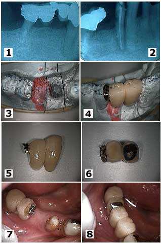
Periapical digital x-rays show how to attach a one porcelain metal dental bridge to another porcelain metal dental bridge in the middle of a pontic tooth. 1) This elderly patient developed distal root caries under a fixed teeth bridge and a root canal infection on a lower first premolar tooth. Note that the distal molar had previously been hemisected. 2) Following removal of the teeth bridge section and root canal treatment. 3) – 4) A two and a half unit dental bridge fabricated and seated on the working model. 5) – 6) Two photo views of the two and a half unit teeth bridge. The distal half unit has been fabricated with a female semi-precision attachment. 7) Intra oral photo. Note how the metal framework of the pontic was prepared into a “T” shape to help lock in the bridge upon cementation. 8) The final dental bridge.
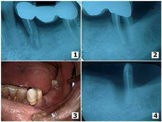
Digital periapical dental x-ray series show diagnosis of the trauma from a fall in an elderly male. The accident caused a broken tooth root that was the mesial abutment of a three teeth bridge. A furcation involvement can be seen in the xray of the distal molar abutment tooth. 1) – 2) The pre-operative radiographs x-rays. 3) The second premolar tooth was extracted and the first premolar was used as the mesial abutment of a new four teeth fixed dental bridge. 4) The second molar tooth was hemisected because of deep probing depths around the mesial root and the mesial root was extracted. The distal root was used as the distal abutment of the new teeth bridge. Bone fill can be seen in the periapical x-ray around the mesial surface of the distal root; a cribriform plate and periodontal ligament space can be seen developing.
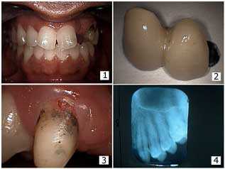
Dental x-ray taken during repair of a fixed porcelain teeth bridge. 1) – 2) Patient had a two-unit, two teeth dental bridge on an upper canine abutment tooth, a lateral incisor pontic and palatal wing attached to the distal of the central incisor tooth. The patient presented with the bridge out. 3) Initially it seemed that there was external resorption on the facial surface of the canine tooth near the gingival margin. 4) The starting periapical xray fortunately did not show resorption so this dental bridge repair was made without further complication.
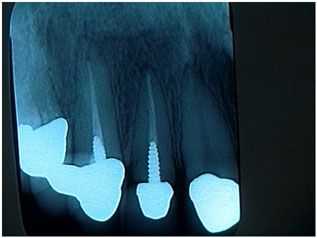
Dental diagnosis based upon a dental x-ray. This nice new patient presented for an emergency visit for what she believed would be a quick recementation of a loose upper lateral tooth crown. This periapical dental xray shows the many recent dental crowns and root canal treatments that were recently completed by her local dentist near her home. She indicated that she had a good relationship with him.
I chose not to treat this patient and instead recommended that she go back to her current dentist for dental care. 1) It appeared that the loose dental crown was completely seated on the post and core instead of being seated on a minimum of two millimeters of sound tooth structure. 2) The post preparation was short and should have been nearly double its length for strength and retention. 3) The post was prefabricated and not cast; this will also decrease its strength and retention.
I did not want to take out the loose tooth crown and then find that I also had a loose post and core in my hand. Proper care might indicate a new, longer cast post and core, possible crown lengthening gum surgery and a new tooth crown. Crown lengthening gum surgery could likely affect the dental crown margins on all the other new anterior teeth crowns.
Once you touch it, you own the problem. Both the dentist and patient need to agree in writing with what might happen, and then what might need to be done, if an “easy” repair turns out to be quite a big problem. This philosophy is both fair and appropriate for the patient and will save a lot of young dentists from having a major, unexpected problem. All things considered it was not worth getting started. The patient was appreciative of my assessment and was happy to return to her dentist for the repair. I hear a lot of patient stories of dentists who begin dental treatment without first taking a dental xray. It is a very poor idea.
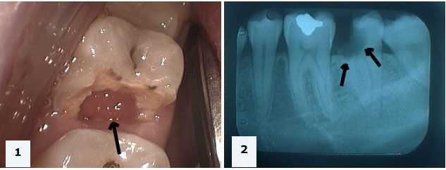
This photo and digital periapical xray shows a hopeless tooth that needs extraction. 1) Photo of tooth #18 showing gum tissue growing into the tooth gap space previously occupied by tooth structure. 2) The dental x-ray shows extensive tooth decay extending below the level of the gums and the jaw bone. Without taking a dental xray a dentist might try to save this tooth but it will ultimately prove to be a bad decision. Dental radiographs x-rays are an important part of dental diagnosis. If a new patient wants dental treatment but refuses to take an xray you should refer them elsewhere.