Photos on removal of porcelain crown and bridge by cutting created in our General Dentistry office.
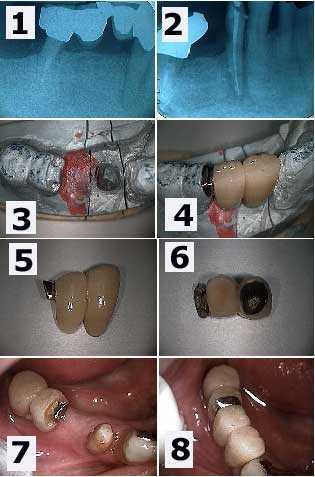
Tooth root cavity and root canal infection around a lower right first premolar. 1) A large radiolucency is seen in this xray. Note the distal abutment is the distal root of a first molar following hemisection. 2) Following root canal therapy. The cervical decay – cavity – can now be seen more easily. 3) The working model showing the preparation of the mesial part of the distal two-unit bridge. This preparation will allow rigid connection to the new anterior bridge being made. 4) The new bridge section in place on the working model. 5) & 6) Two photos of the new dental bridge showing the distal attachment. 7) An intra oral view which mirrors the working model view seen in 3). 8) The final result. This patient was content to see the gold occlusal rest or porcelain could have been added to it.
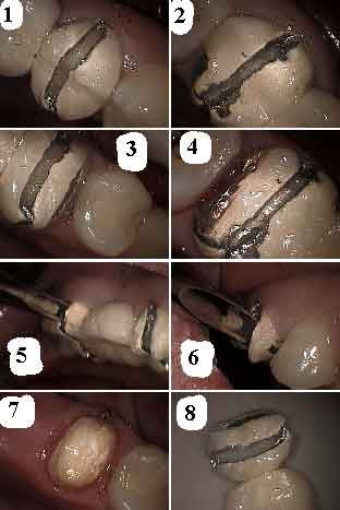
Removal by cutting, sectioning of a three tooth dental bridge. How to pictures:
1) Cut through mid buccal and occlusal with diamond through porcelain and then use a steel bur; e.g. 700xl through metal premolar tooth.
2) Same concept molar tooth.
3) Interproximal reduction premolar tooth.
4) Interproximal reduction molar tooth.
5) Place elevator in slot and rotate gently molar tooth.
6) Same concept premolar tooth — the interproximal reduction gives room for the part of the bridge being rotated off and protects adjacent teeth.
7) Exposed abutment teeth are intact.
8) Bridge off in one piece.
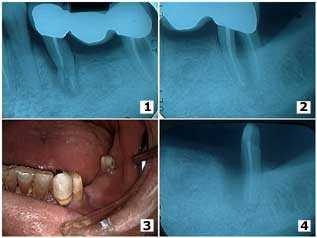
Trauma to a lower left bridge after falling down and hitting the jaw. 1) The initial radiograph showing the three-unit bridge and severe damage to the second premolar. 2) The second molar showing a furcation involvement and thickening of the PDL space around the mesial root. The second premolar and the mesial root of the second molar were extracted. 3) Intra mouth view following healing. The patient was informed regarding the long span and declined implants. He wanted a new fixed bridge anyway. 4) Radiographic healing of the distal root of the second molar after six weeks. The lesson here is the value of salvaging individual roots of molars.
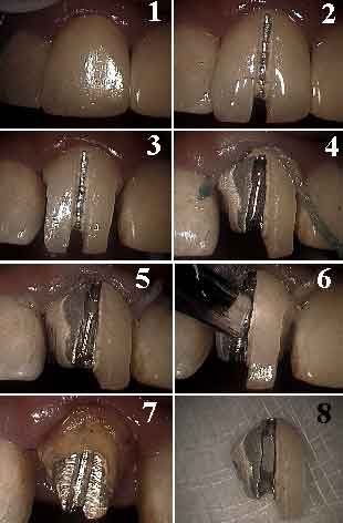
How to picture sequence for gentle removal of an upper front dental crown.
1) Crown tooth #9.
2) Cut through porcelain with barrel-shaped diamond at mid-labial.
3) Cut away interproximal porcelain contacts with flame-shaped diamond.
4) Pack cord before cutting crown
margin to protect gingiva.
5) Crown margin cut.
6) Elevator placed to gently loosen cementation, don’t force it — the interproximal reduction gives room for the part of the crown being rotated off and protects adjacent teeth.
7) Tooth exposed. Differentiate metal in crown from the underlying core.
8) Crown removed in one piece.
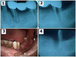
Tooth root fracture of the mesial abutment of a lower left dental bridge and furcation involvement in the distal molar tooth abutment. 1) – 2) Starting x-rays. 3) The second premolar was removed and the first premolar was used as the mesial abutment of a new four-unit bridge. 4) The second molar was hemisected because of deep probing depths around the mesial root and the mesial root was extracted. The distal root was used as the distal abutment of the new dental bridge. Bone fill can be seen around the mesial surface of the distal root; a cribriform plate and periodontal ligament space can be seen developing.