Photos on silver fillings are also called dental amalgam created in our General Dentistry office.
How to drill remove a tooth filling amalgam fillings.
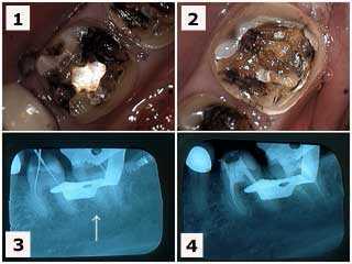
1) & 2) Tooth #’s 19 & 18 following removal of large old silver fillings in a 38 year-old female patient who had been experiencing pain. 3) X-ray of endodontic file lengths. Note that the calcification in the mesial root of #18 initially prevented instrumentation. 4) Final endodontic obturation showing that the mesial root of #18 was located and treated. Chronic inflammation in the dental pulp due to the presence of large, old fillings can increase the difficulty of root canal therapy. Earlier root canal therapy should be considered in these situations.
1) & 2) Tooth #’s 19 & 18 following removal of large old silver fillings in a 38 year-old female patient who had been experiencing pain. 3) X-ray of endodontic file lengths. Note that the calcification in the mesial root of #18 initially prevented instrumentation. 4) Final endodontic obturation showing that the mesial root of #18 was located and treated. Chronic inflammation in the dental pulp due to the presence of large, old fillings can increase the difficulty of root canal therapy. Earlier root canal therapy should be considered in these situations.
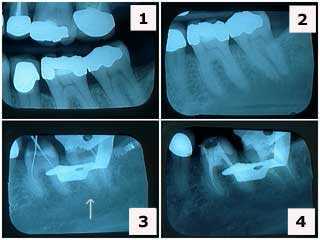
1) & 2) X-rays of tooth #’s 19 & 18 showing large, old silver fillings in a 38 year-old female patient who had been experiencing tooth pain. Note the decreased size of the nerve pulp chambers in these teeth. 3) X-ray of the root canal endodontic file lengths. Note that the tooth calcification in the mesial root of tooth #18 initially prevented endodontist instrumentation. 4) Final endodontic obturation – root canal filling – showing that the mesial root of #18 was located and treated. Chronic inflammation in the dental pulp due to the presence of large, old amalgam fillings can increase the difficulty of root canal therapy. Earlier root canal therapy should be considered in these situations.
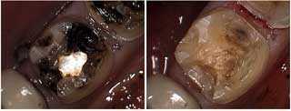
Tooth decay cavity and stain removal following drilling away a large, old silver tooth filling.
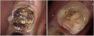
These pictures show tooth decay – dental caries – and stain removal following drilling away of a large, old amalgam dental filling.
Xrays and discussion of a tooth filling tooth – dental amalgam silver filling.
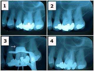
1) & 2) X-rays of tooth #14 showing a large, old tooth filling in a patient who had been experiencing tooth pain. Note the decreased size of the nerve pulp chamber in this tooth. 3) X-ray of endodontic file lengths. Note that the calcification in the mesial root of #14 initially prevented root canal instrumentation. 4) Final endodontic obturation filling showing that the mesial tooth root of #14 was located and treated. Chronic inflammation in the dental pulp due to the presence of large, old amalgam fillings can increase the difficulty of root canal therapy. Earlier root canal therapy should be considered in these situations.
1) & 2) X-rays of tooth #14 showing a large, old tooth filling in a patient who had been experiencing tooth pain. Note the decreased size of the nerve pulp chamber in this tooth. 3) X-ray of endodontic file lengths. Note that the calcification in the mesial root of #14 initially prevented root canal instrumentation. 4) Final endodontic obturation filling showing that the mesial tooth root of #14 was located and treated. Chronic inflammation in the dental pulp due to the presence of large, old amalgam fillings can increase the difficulty of root canal therapy. Earlier root canal therapy should be considered in these situations.