Photos on solder transfer crown and bridge metal frame work created in our General Dentistry office.
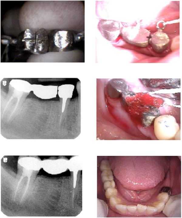
Solder transfer technique for a three unit (three teeth) porcelain to metal fixed bridge. Using a thin carborundum disk a “plus” pattern is drilled into the proximal surfaces of both sides of the framework to be soldered. This helps to tightly hold the GC resin. Note the Kaitlyn loop in the top right photo. The middle left x-ray shows the separate seated copings. GC resin is then applied and the next x-ray shows the seated frame after it was soldered. The final porcelain is later added and seen to the left of the tongue in the last photo.
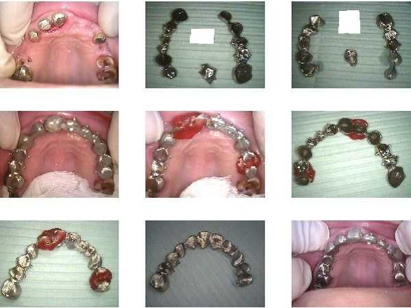
How to technique: solder transfer of a large metal frame in a full-arch fixed porcelain and metal bridge. The top row of photos show the prepped teeth and the metal framework in three sections. The middle row of photos show the three pieces tried on the teeth and then after placement of GC resin. The last two photos in the bottom row of photos show the frame after it was soldered and seated on the teeth to confirm it does not rock. The porcelain should not be added to the metal frame until after this step.
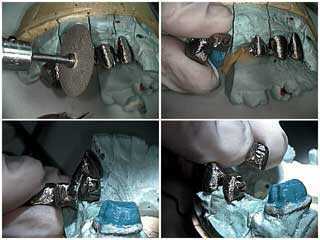
How to pictures for sectioning the metal framework of a five unit fixed porcelain-metal dental bridge. 1) 7/8″ “Veri-thin discs” from Keystone used to section framework which exhibited rocking upon seating. When sectioning, close one eye (wear goggles) and have the disc make exactly a 90 degree contact with the metal to prevent the disc from breaking. 2) In this photo the dental framework is now in two sections and each individual section no longer rocks. 3) and 4) Retention grooves are cut into the proximal sections of the framework to add mechanical retention to Duralay used during the solder transfer step.
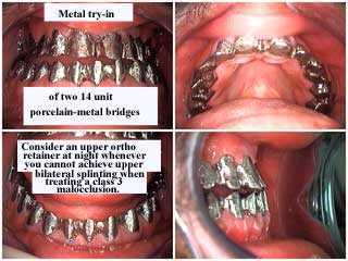
Metal Framework Try-in of a maxillary and mandibular dental bridge. Correction of Under bite, lower jaw in front of upper jaw, and retained baby teeth. Treatment time was 16 days.
Two 14 unit porcelain-metal bridges, were used to increase vertical dimention and “jump” the cross bite. This patient definitely decided against orthognathic treatment.
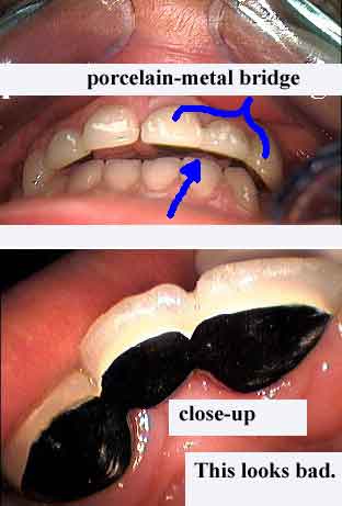
Avoid showing palatal metal when making porcelain-metal dental bridges because it looks bad! Sometimes it may be unavoidable if the patient has a very deep bite and short teeth.
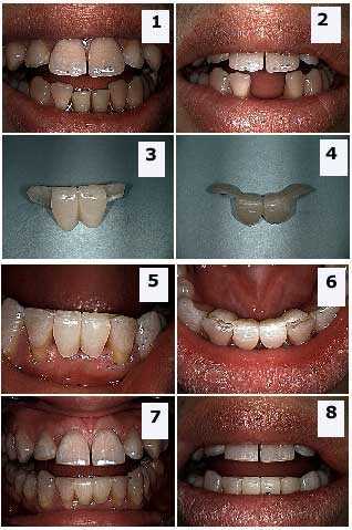
Placement of a Maryland Bridge to replace two missing lower central incisors. 1) Pre-op photo with the patient’s removal partial denture in place. 2) Pre-op photo with the denture removed. 3) – 4) Two extra oral photos of the Maryland bridge. 5) – 8) Four post-op pictures following dental bonding of the bridge to the lateral incisor teeth. These teeth bridges can work well in the anterior when a patient is willing to place less force upon them.