Photos on trauma teeth injury from falling accidents or sports created in our General Dentistry office.
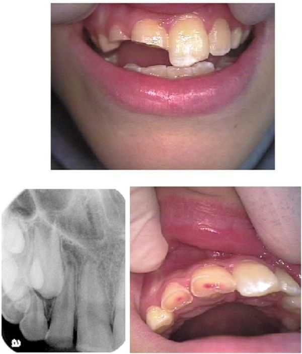
Trauma to two upper front teeth #7 & 8 in a 10 year old boy. Treatment sequence: 1) rule out neurologic injury and immediately refer to ER or neurologist if necessary, 2) rule out facial trauma with PAN and Oral Surgeon consultation, 3) Pedodontist consultation – apexification of #7 & 8 is very important, 4) Endodontist consultation – apexification of #7 & 8 is very important (a partial pulpotomy using MTA was performed), 5) Then consider restorative dentistry.
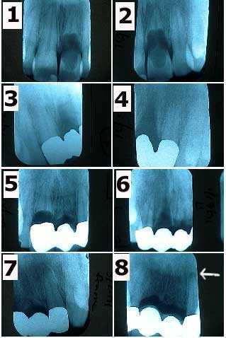
An extreme case of internal resorption occurring within several teeth of a 30 year-old male over ten years. Radiographic series. 1) & 2) The patient related a history of accidental injury from falling to the upper front teeth when these xrays were taken in 1/91. Tooth #9 is undergoing severe internal resorption. 3) & 4) X-rays of the framework of a three teeth bridge in 3/91 following tooth extraction of #9. 5) Tooth #8 exhibiting severe internal resorption in 5/93. 6) & 7) X-rays of the framework of a four teeth bridge in 7/93 following tooth extraction of #8. 8) X-rays of the abutments #7 & 10 in 4/00 shows a periapical radiolucency appearing around #10. The patient was informed of the need for root canal therapy. Xrays in 5/95 & 12/97 had not showed pathologic changes.
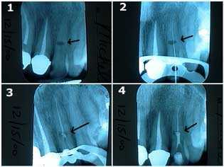
How to treat internal resorption in an upper left lateral incisor tooth. X-ray series. This 40-year-old female related a history of injury to her front teeth after falling during a horseback riding accident. 1) The oval-shaped radiolucency in the middle of the tooth length is the site of the internal resorption. The patient was informed of the guarded long-term prognosis of the tooth. 2) & 3) Traditional root canal therapy was first performed. The canal space was cleaned and shaped, sterilized with NaOCl, and then filled with gutta percha up to a point apical to the internal resorption. 4) MTA, Mineral Trioxide Aggregate, was then placed into the area of the internal resorption and coronal to it. Keeping the MTA moist will give more working time for condensation of material.
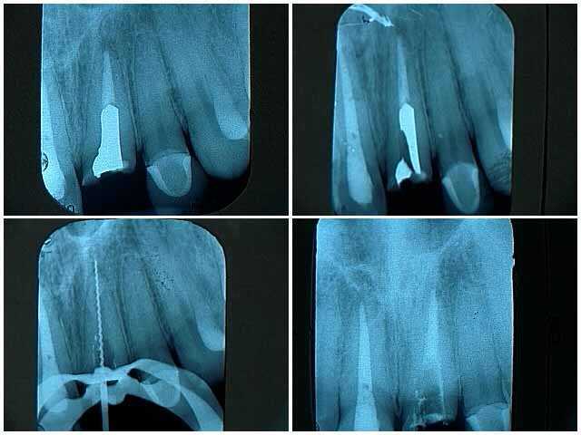
This x-ray series shows the treatment of an upper front central incisor tooth that broke off at the gum line after an injury sustained from falling. ALSO SEE SECTION: OCCLUSION
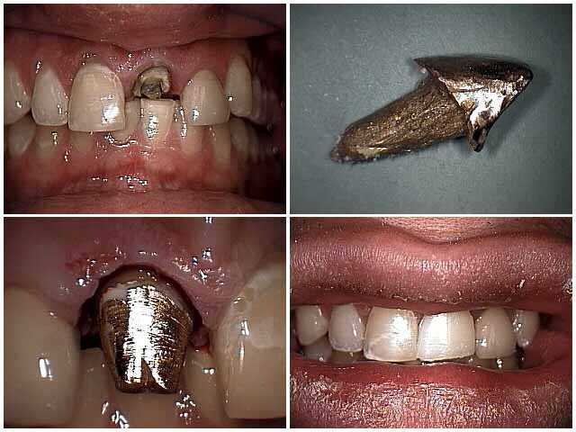
Treatment photos of an upper central incisor tooth broken off at the gum line due to a falling injury. These pictures show 1) the tooth after root canal, 2) the cast gold post and core, 3) the gold post in the tooth after placement of dental cement and 4) the final tooth crown in place.
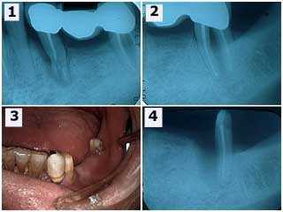
Traumatic injury to a lower left dental bridge following a falling accident. 1) The initial x-ray radiograph shows the three teeth bridge and severe damage to the second premolar tooth. 2) The second molar shows a furcation involvement and thickening of the periodontal ligament space around the mesial root. The second premolar and the mesial root of the second molar were extracted. 3) Intra-oral photo following wound healing. The patient wanted a new fixed bridge despite the long span. 4) This xray shows healing around the distal root of the second molar six weeks later.
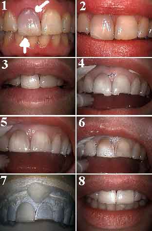
Treatment of trauma to a front tooth from a falling accident. 1) In this Before photo, tooth #8 shows a burst blood vessel through enamel and deep cracks. 2) Following root canal therapy and “walking” internal bleach. 3) Following external office whitening of all teeth. 4) Beginning incisal reduction for porcelain veneers. The patient wanted to eliminate facial tooth bonding on #9 and deep crack lines. 5) Beginning facial reduction using depth cuts. 6) Final preparation for porcelain veneers. 7) Porcelain veneers on the master working model. 8) After photo with the porcelain teeth veneers cemented. ALSO SEE SECTION: OCCLUSION