Before and after photos on gingival margins are the gum line around teeth performed in our Gum Disease Treatment office.
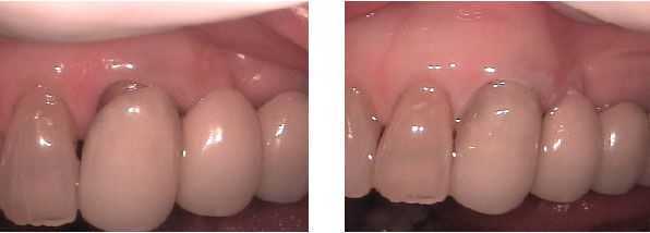
Gingival recession (receded gums) around the gingival margin of a dental bridge shows dark tooth root. This before and after photo shows the benefit of cosmetic teeth bonding without requiring replacement of the bridge.
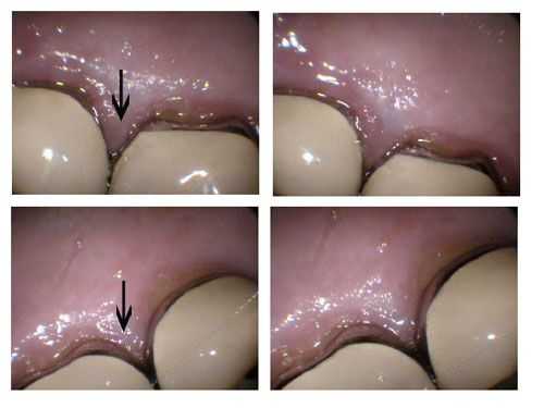
The top photos show inflammation of the the gingival margins – gums – between two upper right dental crowns. The top arrow points to a purple, inflamed gingival papilla – gum point – and gingival embrasure. The bottom pictures show two upper left dental crowns in the same patient that are not inflamed. The bottom arrow points to pink, non-inflamed gum tissue.
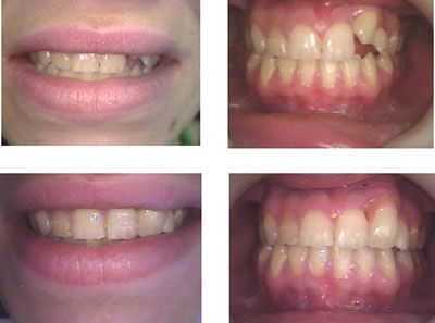
Gingival margins – the gum line – and Cosmetic Dentistry. One tooth bonding and one porcelain veneer. Treatment time: 2 visits. The patient refused teeth braces to bring the tooth down. The Veneer had pink porcelain around the gingival margin gum line to hide its relative height.
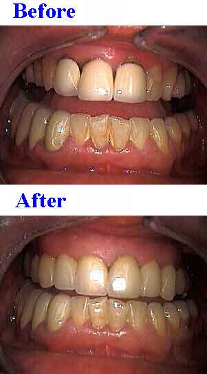
Gingival margins – the gum line and poorly fitting dental crowns. Before and after pictures. The dental crowns in the upper photo were replaced with porcelain tooth crowns seen in the lower photo. Notice after treatment the improved appearance at the gums and the reestablished gingival papillae and gingival embrasures.
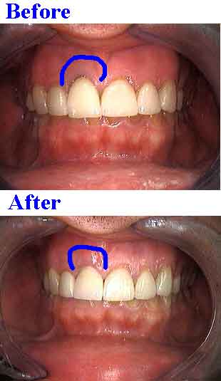
Removing gingival margin darkness – black gums – around a front tooth covered with a porcelain fused to metal dental crown. Minor gum surgery was performed to provide root coverage and a new all-porcelain dental crown was placed.
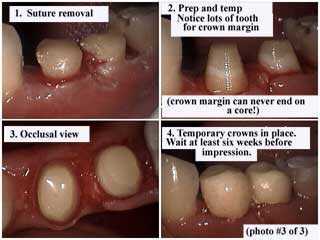
Gingival margins – the gum line after crown lengthening gum surgery, suture removal and dental crown preparation – drilling. Notice a lot of tooth structure has been obtained for the dental crown margin. Dental crown margins can NEVER end on a core. After the temporary dental crown is placed, wait at least 6 weeks before taking an impression.
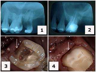
Where to end a dental crown finish line relative to the gums – the gingival margin. Treatment of a a maxillary first molar with severe tooth decay cavity. 1) Initial x-ray. 2) X-ray following tooth extraction of #15 and root canal therapy on tooth #14. Note the gingival extent of the tooth decay – dental caries. 3) Tooth preparation following root canal therapy and the removal of the distal tooth decay. 4) Final tooth preparation of the composite resin crown buildup. Note how the distal tooth treatment extends at the gums – at the gingival margin – beyond the composite resin to solid tooth structure.
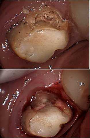
Drilling removal of tooth decay cavity around a molar to establish the expected location of the gingival margin of the dental crown. This should be performed before referring the patient for crown lengthening periodontal gum surgery so the periodontist knows how much osseous bone reduction is necessary.
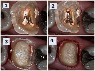
How to pictures and explanation. Crown buildup following root canal therapy in a molar tooth. 1) Following root canal therapy. 2) Tooth preparation for crown buildup. Note preparation into the root canal orifices creates mechanical retention of the core. 3) The Initial shoulder preparation of the dental crown margin. 4) Bevel placement of the dental crown margin. This tooth needs crown lengthening periodontal gum surgery to move the gingival margin apically. Not doing this will mean the tooth crown margin will invade the biologic width resulting in chronic periodontal inflammation.
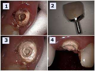
Failure of a maxillary anterior porcelain dental crown. 1) Severe dental caries tooth decay. 2) The failed porcelain tooth crown with the prefabricated post still in it. 3) and 4) Tooth preparation drilling to determine the necessity for crown lengthening gum surgery. The dental crown margin should end on two millimeters of healthy tooth structure. This will allow for a retentive dental crown and healthy gums at the gingival margin.
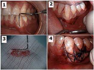
Changing the location of gingival margins – the gum line with a free gingival graft – gum surgery. Free gingival graft for a lower left canine tooth. 1) Periodontal probe in the gingival sulcus showing the mucogingival defect. 2) Initial sub-sulcular surgical incision. 3) The gingival tissue harvested from the palate that will be used for the gum graft. 4) The gum graft sutured in place. Photo #1 of 4.
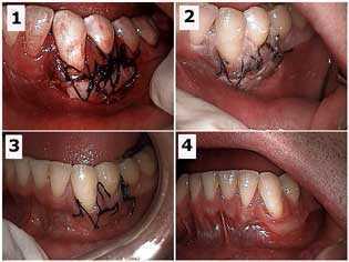
Changing the location of gingival margins – the gum line with a free gingival graft – gum surgery. Free gingival graft of a lower left canine tooth. Healing of the recipient site. 1) Initial surgical site. 2) One week post-op. 3) Two weeks post-op. 4) Seven weeks post-op. Photo #2 of 4.
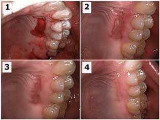
Changing the location of gingival margins – the gum line with a free gingival graft – gum surgery. Free gingival graft of a lower left canine tooth. Pictures show healing of the palatal donor site. 1) Initial gum surgical site. 2) One week post-op. 3) Two weeks post-op. 4) Seven weeks post-op. Photo #3 of 4.
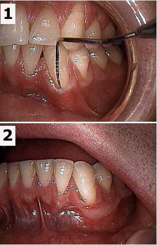
Changing the location of gums with a free gingival graft – gum surgery. Before and after pictures of a free gingival graft of a lower left canine tooth. 1) Periodontal probe in the gingival sulcus shows the mucogingival defect. 2) Seven weeks post-op. Photo 4 of 4.