Before and after photos on gingivectomy around crowns bridges and teeth performed in our Gum Disease Treatment office.
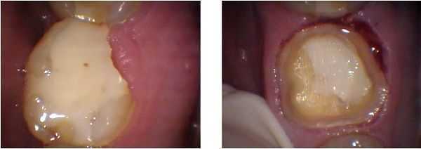
Gingivectomy performed during a crown preparation. The distal lingual cusp of this tooth had fractured and the patient ignored it for a while. This caused the overgrowth of the gum in this area.
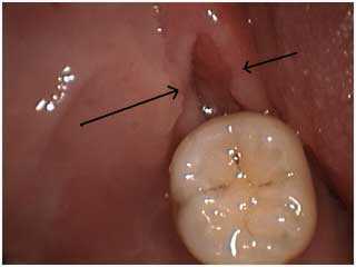
Distal wedge procedure – one week post op. This periodontal gum surgery is performed to remove gum tissue – the pericorium – that may grow over the biting surface of the lower back molar. The pericorium in this patient had occasionally resulted in tooth pain infection and required antibiotics.
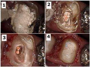
Crown buildup and periodontal gum surgery on a maxillary first molar after removal of tooth decay and root canal therapy. 1) Temporary filling material in the tooth following root canal therapy. 2) Gutta percha exposed in the pulp chamber after removal of the temporary dental filling. Tooth decay is still present. 3) Tooth cavity removal and preparation into the root canal orifices to aid in mechanical retention of the dental bonding composite resin core. 4) Final drilling preparation after gum surgery in the distal section of the tooth.
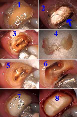
Crown buildup with gingivectomy – GV.
1) Old composite dental filling.
2) Composite removed, recurrent decay is visible.
3) Pulp chamber with tooth decay cavity still present.
4) Old composite came out in one piece; it was not bonded.
5) Tooth decay removed and prep into canal orifices for bonding retention.
6) Depth of drilling preparation into pulp chamber.
7) Dental bonding is in place before tooth preparation.
8) Tooth preparation and gingivectomy; note the occlusal retention groove.
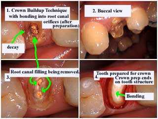
Crown buildup technique with dental bonding into root canal orifices after preparation. Notice dental crown preparation ends on tooth structure.
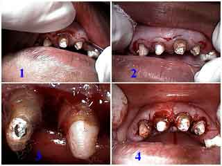
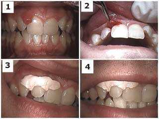
Short clinical teeth crowns that require crown lengthening gum surgery to increase retention for the maxillary anterior dental bridge. 1) and 2) A three millimeter submarginal incision was made around all the teeth. 3) The gingival collar was removed including the interproximal tissue. 4) The flap was sutured against the underlying bone.
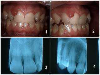
Pyogenic granuloma on the papilla – gum point – between two teeth of a 40-year-old male. The x-rays radiographs revealed no pathology. The soft tissue was firm and did not respond to periodontal scaling. It did not hurt and did not bleed upon probing. Initial diagnosis: irritation fibroma. The tissue was sent for biopsy; the pathology report indicated it was a pyogenic granuloma. Photo 1 of 3.
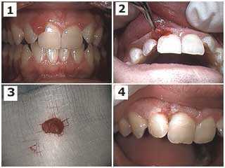
Pyogenic granuloma on the papilla between two teeth of a 40-year-old male. The x-rays radiographs did not reveal pathology. The gum tissue was firm and did not respond to root planing and scaling. There was not associated pain and it did not bleed upon probing. Treatment: surgical excision. The tissue was sent for biopsy; the pathology report indicated it was a pyogenic granuloma. Photo 2 of 3.
Pyogenic granuloma on the papilla between two teeth of a 40-year-old male. The radiographs revealed no pathology. The gum tissue was firm and did not respond to scaling and root planing. It did not hurt and did not bleed upon probing. Treatment: surgical site after excision and placement of Coe-pak periodontal wound dressing. The tissue was sent for biopsy; the pathology report indicated it was a pyogenic granuloma. Photo 3 of 3.
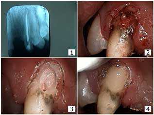
Gingivectomy with Dental Bridge repair. The patient wanted to try to save his bridge. 1) X-ray after root canal therapy. The root canal itself did not seem subject to external resorption. 2) Gingivectomy GV. Note the still unusual look of the facial tooth decay. 3) Following tooth decay cavity removal. Note the communication to the gutta percha. 4) The dental bonding restoration. Photo #1 of 2.
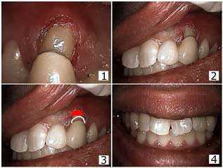
Gingivectomy with Dental Bridge repair. The patient wanted to save his dental bridge. 1) Fitting the teeth bridge over the tooth bonding repair. 2) This teeth bridge was cemented with resin cement. The mesial wing was also bonded to the distal of the lateral incisor tooth. 3) The gingivectomy was chosen over a gum surgery flap because the patient had a low lip line and so the development of a pseudopocket with a flap was avoided. The red indicates where the gum line could have been. 4) The final result. The patient is informed of the need for bridge replacement when finances allow. Photo #2 of 2.