Before and after photos on gum grafts bone grafts and sinus elevation performed in our Gum Disease Treatment office.
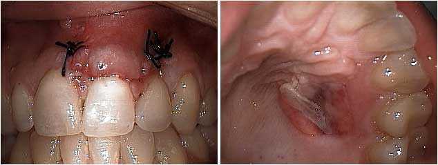
Free gingival graft gum surgery after three days. The upper front central incisor tooth exhibited a lot of gingival gum recession that bothered the patient cosmetically. 1) The gum graft is shown after being sutured in place. These black sutures will be removed in about ten days. 2) The palate of the same patient showing the donor site for the gum graft. A gingival graft typically involves removing a small amount of palatal gingiva and sewing it where it is needed.
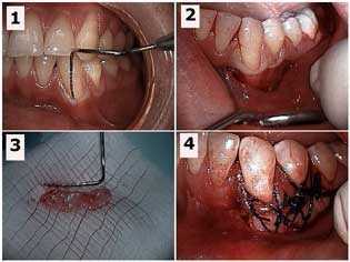
How to Pictures: free gingival graft gum surgery for a mandibular canine tooth. 1) Periodontal probe in the gingival sulcus showing the mucogingival defect. 2) Initial sub-sulcular incision. 3) The gingival gum tissue harvested from the palate that will be used for the graft. 4) The graft sutured in place. Photo #1 of 4.
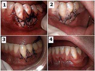
How to Pictures: free gingival graft gum surgery of a mandibular canine tooth. Healing of the recipient site. 1) Initial surgical site. 2) One week post-op. 3) Two weeks post-op. 4) Seven weeks post-op. Photo #2 of 4.
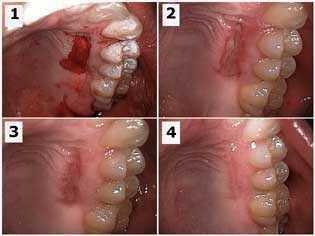
Free gingival graft gum surgery of a mandibular canine tooth. Healing of the palatal donor site. 1) Initial surgical site. 2) One week post-op. 3) Two weeks post-op. 4) Seven weeks post-op. Photo #3 of 4.
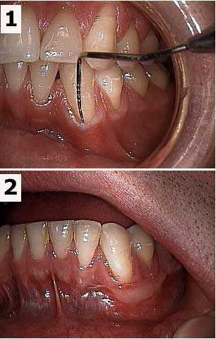
Before and after pictures of a free gingival graft gum surgery of a mandibular canine tooth. 1) Periodontal probe in the gingival sulcus shows the mucogingival defect. 2) Seven weeks post-op. Photo #4 of 4.
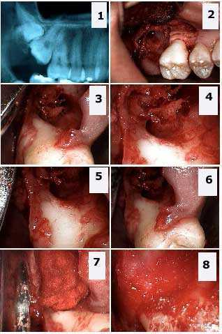
Extraction of a double tooth impaction: upper right third wisdom and second molars. 1) Xray shows the double tooth impaction. 2) – 6) Different images show the large osseous bony defect and the significant exposure of the distal furcation of the first molar. 7) – 8) Packing the defect with freeze-dried bone and gelfoam.
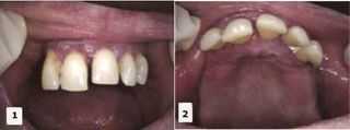
Dental implant oral surgery with sinus elevation and bone graft pictures. A 64 year-old female presented with five very loose anterior maxillary teeth and missing all posterior maxillary teeth. She wanted a fixed teeth bridge. The patient had six dental implants placed in the mandible (lower jaw) with fixed caps about four years earlier. The panorex x-ray showed an atrophic maxilla with 4mm of bone, which required a bone graft and sinus lift for dental implants. The agreed treatment plan consisted of extractions of all anterior teeth, sinus lifts with platelet-rich plasma (PRP), and simultaneous implant placement. Part 1 of 5.
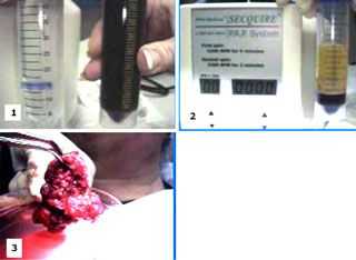
Dental implant oral surgery with sinus elevation and bone graft how to pictures. 1) A Platelet Concentration Specialist obtained the patients’ own blood (60 cc) and 2) centrifuged it to obtain 6-8 cc of platelet-rich plasma (PRP) and 10 cc of platelet-poor plasma (fibrin glue). 3) The PRP was mixed with the patients’ bone and freeze-dried bone, and then activated with thrombin. This mixture was then used as graft material for the sinus lift elevation in and around the implants. Part 2 of 5.
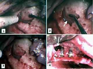
Dental implant oral surgery with sinus elevation and bone graft how to pictures. 1) – 3) A sinus lift elevation was performed by opening a small “window” into the sinus without perforating the Schneiderian Membrane. The window was gently elevated along its superior fracture line and the membrane peeled off of the sinus floor, creating a new, more superior sinus floor. 4) The dental implants were carefully placed to make sure they were stable. Notice that the width of the alveolar ridge was inadequate for the dental implants necessitating the PRP graft. The most distal two implants can be seen extending vertically into the elevated sinus. This entire area was covered with the PRP graft. Part 3 of 5.
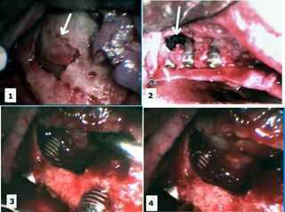
Dental implant oral surgery with sinus elevation and bone graft how to pictures. 1) Sinus elevation. 2) Implants placed in the elevated sinus. 3) – 4) Close-up of the implant in the sinus with the new floor of sinus superiorly positioned. This entire area was covered with PRP graft. Part 4 of 5.
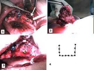
Dental implant oral surgery with sinus elevation and bone graft how to pictures. 1) Palatal photo of the dental implant site showing the area of the sinus lift elevation. 2) – 3) The graft (PRP + patients’ bone + freeze-dried bone) material was then placed in the sinus cavity and around the dental implants. The wound was then closed primarily and the bone graft was allowed to consolidate for 9 months. 4) Pattern of preparation in bone for sinus elevation. Part 5 of 5.