Before and after photos on horizontal bone loss from gum disease in x-rays performed in our Gum Disease Treatment office.
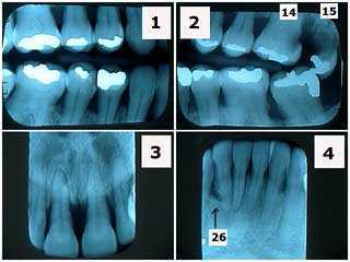
X-ray series showing severe periodontal gum disease in a 40-year-old female with dental anxiety. Note the significant horizontal bone loss in the jaw and the lack of radiographic calculus. Tooth #26, adjacent to the vertical bony defect, is probably hopeless. This patient was treated with two rounds of scaling and root planing then periodontal reevaluation. Tooth #15 was extracted and #14 had root canal therapy and a crown.
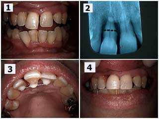
The patient’s chief complaint was that the upper right central incisor (#8) was very loose in her upper jaw and she did not want to lose the tooth. 1) Upper right central incisor (#8) has extruded and moved labially out of the mouth (toward the lip) and a diastema (space) has occurred. 2) Radiograph showing severe bone loss. The black dotted line shows the location for a potential root resection if necessary. Root planing was performed after the splint was placed and the patient will return in three months to reevaluate periodontal healing and the potential need for the root resection. 3) Palatal view of the splint between teeth #’s 7 – 9. 4) Post-op view. Same day. Notice the diastema (space) was closed with bonding to hide the splint and the incisal edge of #8 was reduced.
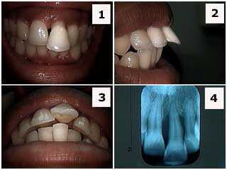
Patient presented with her upper left central incisor flared labially. Extra oral pictures. 1) Front view photo. 2) Side view photo shows the tooth protruded out of the jaw. 3) Occlusal view photo. 4) x-ray, Radiograph. The patient, who was in her twenties, was informed about the severe periodontal condition and that the prognosis for this tooth was guarded at best. A thorough periodontal evaluation was recommended. Orthodontics is contra-indicated here because of the severe periodontal problem. Treatment, if the patient accepts treatment on a tooth with a guarded prognosis, could include: root planing and scaling, open flap debridement if necessary, root canal therapy and crown (for esthetics) and possibly a splint. Treatment, otherwise, could include just scaling and root planing and incisal adjustment. I personally would not rush to extract this tooth though the patient needs to be aware of this possibility.
Horizontal jaw bone loss in a full mouth reconstruction patient. x-ray series.