Before and after photos on mucogingival involvement and attached gingiva performed in our Gum Disease Treatment office.
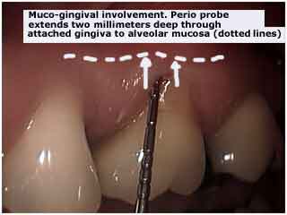
Mucogingival involvement – gingival gum recession- on a maxillary molar tooth. Note the periodontal probe measuring the extent of the gum recession.
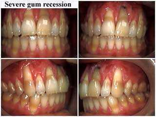
Severe gum recession – receded gums on all teeth in the mouth. Photos of severe mucogingival involvement.
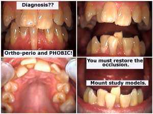
Severe mucogingival involvement – gum recession – on a lower front tooth is seen in these photos of this full mouth reconstruction, dental phobia patient.
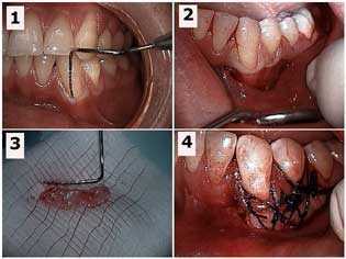
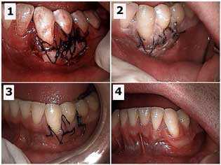
How to treatment pictures of mucogingival involvement – gingival gum recession to the mucogingival junction. These photos show a free gingival gum graft for a lower left canine tooth. 1) Periodontal probe in the gingival sulcus – gum pocket – shows the mucogingival defect. 2) Initial sub-sulcular surgical incision in the gums. 3) The gingival tissue harvested from the palate that will be used for the gum graft. 4) The free gingival graft is sutured in place. Photo #1 of 4.
How to treatment pictures of mucogingival involvement – gingival recession to the mucogingival junction. These photos show healing of the recipient site gums following a free gingival gum graft for a lower left canine tooth. 1) Initial surgical site of the free gingival graft. 2) One week post-op. 3) Two weeks post-op. 4) Seven weeks post-op. Photo #2 of 4.
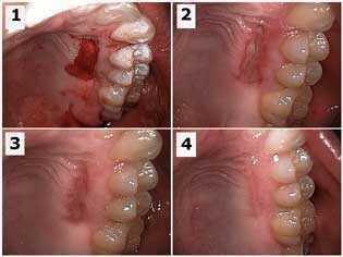
How to treatment pictures of mucogingival involvement – gingival gum recession to the mucogingival junction. These photos show healing of the palatal donor site following a free gingival gum graft for a lower left canine tooth. 1) Initial donor surgical site for the free gingival graft. 2) One week post-op. 3) Two weeks post-op. 4) Seven weeks post-op shows the gums healing. Photo #3 of 4.
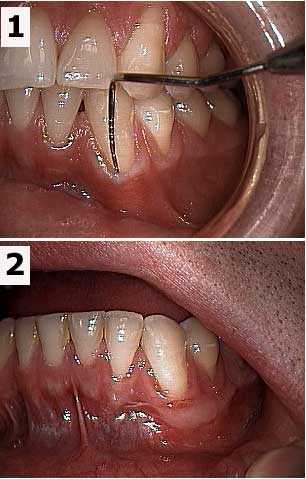
Before and after pictures of a free gingival gum graft of a lower left canine tooth. 1) Periodontal probe in the gingival sulcus shows the muco-gingival defect. 2) Seven weeks post-op. Photo #4 of 4.