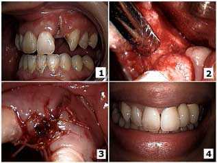
Before and after photos on periodontal regeneration with bone grafting performed in our Gum Disease Treatment office.

Periodontal Generation – how to pictures show oral surgery closure of a buccal dehiscence on an upper lateral incisor tooth. Photo 1) The initial presentation with the lateral incisor tooth root exposed and tooth decay up to the root apex. Photo 2) Surgical exposure of the site after extraction of the tooth root. A full thickness mucoperiosteal gum flap with bucco-lingual vertical releasing surgical incisions was used to obtain primary closure. The tooth socket was curetted and freeze-dried cortical bone graft chips were placed with a Biomend membrane. Photo 3) Primary oral surgery closure was obtained with 3-0 Vicryl sutures. Photo 4) The final result after fabrication of the porcelain-metal fixed dental bridge.