Before and after photos on porcelain dental bridge gums bleed around crowns performed in our Gum Disease Treatment office.
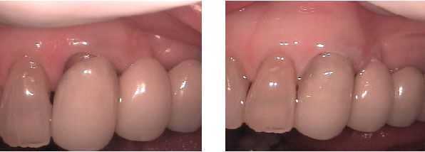
Cosmetic repair of tooth root showing around a bridge margin. Before and after photos following dental bonding.
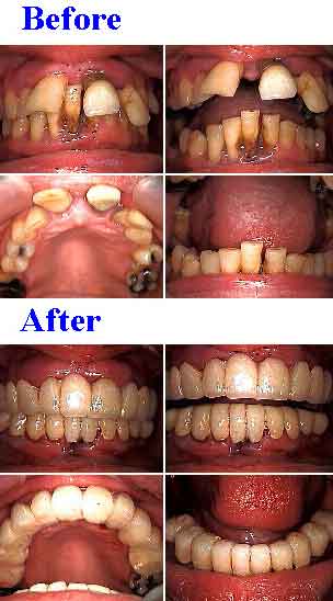
Full Mouth Oral Reconstruction. 20 temporary dental caps (two full jaw provisional teeth bridges), 14 teeth root canals and 6 teeth extractions. Treatment time: one visit.
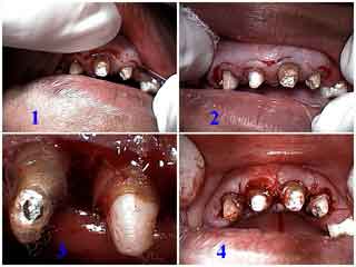
Short clinical teeth crowns that require crown lengthening gum surgery to increase retention for the maxillary anterior dental bridge. Pictures 1) and 2) A three millimeter submarginal incision was made around all the teeth. 3) The gingival collar was removed including the interproximal tissue. 4) The gum flap was sutured against the underlying bone.
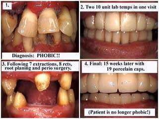
Full Mouth Dental Reconstruction: Diagnosis: Dental Phobia Fear. Treatment: 7 teeth extractions, 8 teeth root canals and composite cores, full mouth scaling root planing and then periodontal gum surgery, 19 units – teeth – of dental bridge work over 8 teeth abutments. Before and after pictures.
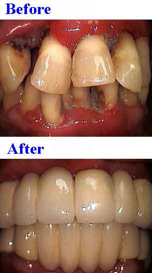
Full Mouth Oral Reconstruction: Treatment: 7 teeth extractions, 8 teeth root canals and composite cores, full mouth root planing and then periodontal surgery, 19 units of bridge work over 8 abutments. Before and after photos.
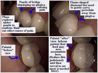
The pontic, false tooth, of this dental bridge was impinging, pushing, on the gingiva, gum, causing pain. How to technique to relieve stress on the gums.
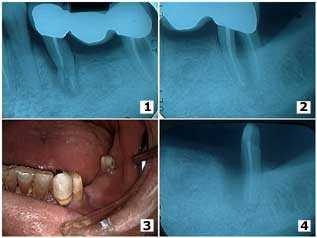
Trauma from a fall in an elderly male resulted in root fracture of the mesial abutment of a three-unit bridge and a furcation involvement in the distal molar abutment. 1) – 2) The pre-operative x-rays. 3) The second premolar was extracted and the first premolar was used as the mesial abutment of a new four-unit bridge. 4) The second molar was hemisected because of deep periodontal probing depths around the mesial tooth root and the mesial root was extracted. The distal root was used as the distal abutment of the new teeth bridge. Jaw bone fill can be seen around the mesial surface of the distal root; a cribriform plate and periodontal ligament space can be seen developing.
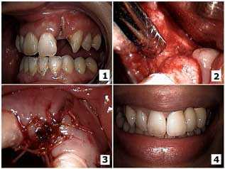
How to pictures. Surgical closure of a buccal dehiscence on an upper lateral incisor. 1) The initial presentation with the lateral incisor root exposed and decayed up to the apex. 2) Surgical exposure of the site after extraction of the tooth root. A full thickness mucoperiosteal flap with bucco-lingual vertical releasing incisions was used to obtain primary closure. The socket was curetted and freeze-dried cortical chips were placed with Biomend membrane. 3) Primary closure was obtained with 3-0 Vicryl sutures. 4) The final result after fabrication of the porcelain-metal fixed dental bridge.