Before and after photos on receded gums treatment includes cosmetic dentistry performed in our Gum Disease Treatment office.
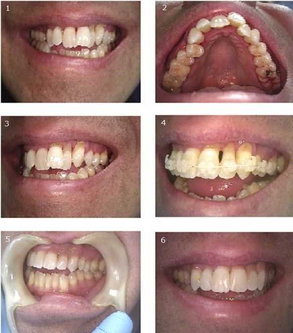
Cosmetic Dental Bonding to hide severe gingival (gum) recession following prior periodontal surgery that completely disregarded the patient’s appearance. #1, 2 & 3) The patient’s chief complaint was the severe gum recession around his upper left teeth and the crooked upper front teeth.
The patient consulted with our Periodontist (gum specialist) and Orthodontist (for braces). He was treated with two rounds of scaling and root planing, conservative, non-surgical gum therapy.The Periodontist suggested limiting movement of the lateral incisor #10 because it had a poor crown to root ratio. The patient was then cleared for limited orthodontics. The patient was put on three month recall exams with the Periodontist during braces treatment. #4) The Orthodontist bonded the upper arch from second premolar to second premolar. These cosmetic braces use white brackets and wire. At patient request the orthodontic treatment plan focused on eight months of braces to align the upper arch only and not to close the open bite. After eight months the braces were removed and a bonded upper lingual retainer was placed.#5 & 6) Cosmetic dentistry options for improving the appearance of the severe gingival gum recession around the upper left teeth focused on either composite resin bonding at the gumline or porcelain laminate veneers with pink gingiva (gums) at the gum line. The patient chose pink composite resin bonding to reduce cost. This procedure was completed in less than one hour.
The patient consulted with our Periodontist (gum specialist) and Orthodontist (for braces). He was treated with two rounds of scaling and root planing, conservative, non-surgical gum therapy.The Periodontist suggested limiting movement of the lateral incisor #10 because it had a poor crown to root ratio. The patient was then cleared for limited orthodontics. The patient was put on three month recall exams with the Periodontist during braces treatment. #4) The Orthodontist bonded the upper arch from second premolar to second premolar. These cosmetic braces use white brackets and wire. At patient request the orthodontic treatment plan focused on eight months of braces to align the upper arch only and not to close the open bite. After eight months the braces were removed and a bonded upper lingual retainer was placed.#5 & 6) Cosmetic dentistry options for improving the appearance of the severe gingival gum recession around the upper left teeth focused on either composite resin bonding at the gumline or porcelain laminate veneers with pink gingiva (gums) at the gum line. The patient chose pink composite resin bonding to reduce cost. This procedure was completed in less than one hour.
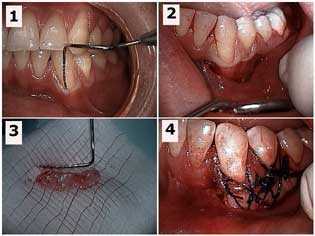
Gum recession – receded gums treatment. How to pictures. Free gingival graft for a lower left canine tooth. 1) Periodontal probe in the gingival sulcus showing the mucogingival defect. 2) Initial sub-sulcular surgical incision. 3) The gingival gum tissue harvested from the palate that will be used for the gum graft. 4) The gum graft sutured in place.
Photo #1 of 4 – see Mucogingival and Recession sections in the left margin for more photos.
Photo #1 of 4 – see Mucogingival and Recession sections in the left margin for more photos.