Before and after photos on the periodontal ligament PDL in health and disease performed in our Gum Disease Treatment office.

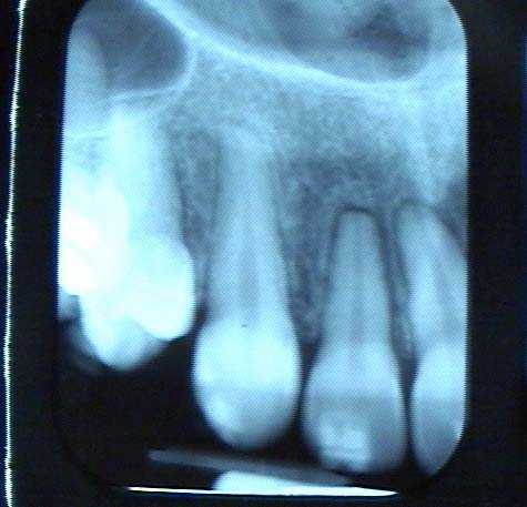
Widening of the periodontal ligament space – PDL space – seen especially around this upper lateral incisor tooth associated with normal orthodontic tooth movement – teeth braces. This will return to normal following the completion of orthodontic tooth movement.

How to pictures for a tooth root resection from under a 10 tooth porcelain fused to metal dental bridge.
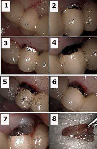
1) Labial photo of an abutment tooth with a combined root canal and gum infection. 2) & 3) Initial preparation – drilling – into the porcelain crown with a diamond bur. 4) Labial photo shows preparation. The metal portion of the teeth bridge is prepared with a steel bur. 5) Palatal photo shows preparation. 6) Labial view showing the tooth root under the porcelain (gutta percha is visible in the tooth root). 7) The tooth root extraction. The occlusal height of this root must first be reduced to allow it to exit. 8) The extracted tooth root.
1) Labial photo of an abutment tooth with a combined root canal and gum infection. 2) & 3) Initial preparation – drilling – into the porcelain crown with a diamond bur. 4) Labial photo shows preparation. The metal portion of the teeth bridge is prepared with a steel bur. 5) Palatal photo shows preparation. 6) Labial view showing the tooth root under the porcelain (gutta percha is visible in the tooth root). 7) The tooth root extraction. The occlusal height of this root must first be reduced to allow it to exit. 8) The extracted tooth root.

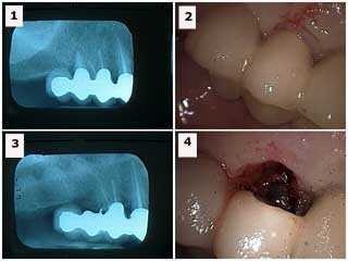
How to pictures for a tooth root resection from under a 10 tooth porcelain fused to metal dental bridge. 1) This x-ray shows the distal tooth abutment with a widened periodontal ligament space. 2) Labial view of the same tooth. 3) Xray after tooth root resection (the metal chad in the area of the extracted abutment was later removed). 4) Labial view following extraction. This area could be filled in with dental bonding after wound healing. The occlusion on the distal cantilever was reduced by making it appear more like a canine on the palatal side. It opposed a lower full arch bridge so that supraeruption was not a concern. Treatment options include: i) sectioning and removal of the distal cantilever, ii) dental implants, or iii) reevaluate over time with the patient informed to reduce function in this area.

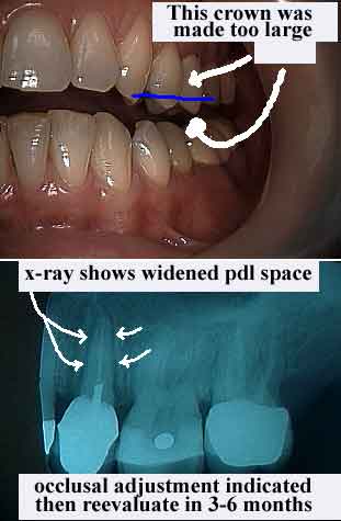
Occlusal Trauma. Note widening of the periodontal ligament space – PDL space- in the xray. This should resolve over time after occlusal adjustment if periodontal pathogens are controlled. Note we refer to the periodontal ligament “space” because in the x-ray we cannot actually see the ligament. We can only see the space where it resides. Early gum inflammation is called gingivitis. Later stage gum disease inflammation is called periodontitis.