Before and after photos on wound healing after oral surgery and periodontal surgery performed in our Gum Disease Treatment office.

Application of Surgicel,an absorbable hemostat made of oxidized regenerated cellulose, to stop bleeding from a minor cut on the tongue that occurred during operative dentistry.
Wound healing following treatment of a painful acute periodontal abscess. 1) This patient presented with a draining fistula in the gums between teeth #’s 7 & 8. Teeth #’s 8 & 9 had acrylic laminate veneers placed about two years ago by another dentist who then performed a gingivectomy last year to attempt to treat the occasional swelling of the gums in this same area. 2) The x-ray of tooth #9 shows a healed apicoectomy from 12 years ago that is not related to the current problem. 3) A gutta percha point was placed in the fistula and x-rayed to see where it lead. It stayed at the coronal gingival margin around the margin of the veneer. The patient was put on tetracycline for a week. 4) Reevaluation two days later showed improvement in symptoms and gingival inflammation. An evaluation and treatment of the bulky veneer margin will occur following resolution of the acute condition. Photo #1 of 2.
Wound healing of a painful acute periodontal gum abscess. A bulky margin of a dental crown or tooth veneer can cause significant inflammation of the gums. Bleeding gums is one symptom. This should be treated before an emergency occurs.
Photo #2 of 2.
Photo #2 of 2.
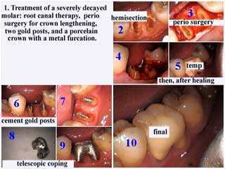
Treatment of a severely decayed molar:
1) root canal therapy. 2) periodontal surgery for crown lengthening. 3) two gold posts were placed after wound healing. 4) porcelain crown with a metal furcation.
1) root canal therapy. 2) periodontal surgery for crown lengthening. 3) two gold posts were placed after wound healing. 4) porcelain crown with a metal furcation.
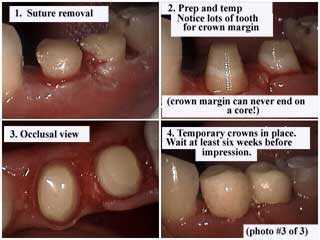
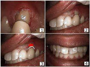
Wait six weeks for wound healing after periodontal crown lengthening gum surgery before taking a final impression for a tooth crown. These photos show after removal of the sutures stitches and the initial crown preparation. Notice lots of tooth for the crown margin — crown margins should not end on a core.
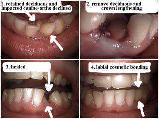
Typically wait 12 weeks for wound healing following tooth extraction before proceeding with comprehensive cosmetic dentistry. The labial surface of the remaining tooth received bonding to close the space after the extraction of the baby tooth.
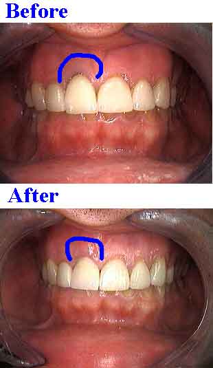
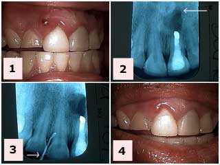
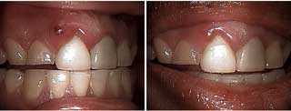
Removing “darkness” around a front tooth. Minor gum surgery and new porcelain dental crown. Wait six weeks for wound healing before completing final cosmetic dentistry unless a laser is used.
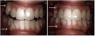
Cosmetic dentistry treatment of upper and lower right canines. The patient declined orthodontics. The upper and lower canines were extracted and the distal of the laterals and the mesial of the first premolars were bonded to close the gap spaces. The second photo is one week following extraction. The bonding was placed before the teeth were extracted so the patient never had to show the space between her teeth. It should be expected that additional cosmetic teeth bonding will be necessary near the gums as the wounds heal over the next 12 weeks. Photo #1 of 2.
Cosmetic dentistry treatment of upper and lower right canines. The patient declined orthodontics. The upper and lower canines were extracted and the distal of the laterals and the mesial of the first premolars were bonded to close the gap spaces. The second photo is one week following extraction. The bonding was placed before the teeth were extracted so the patient never had to show the space between her teeth. It should be expected that additional cosmetic teeth bonding will be necessary near the gums as the wounds heal over the next 12 weeks. Photo #2 of 2.
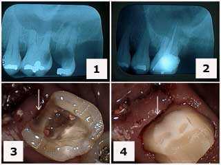
Treatment of a severely decayed upper left first molar #14. 1) Initial radiograph. 2) X-ray following extraction of tooth #15 and root canal therapy on tooth #14. Note the gingival extent of the tooth decay. An ideal time for tooth decay removal is during wound healing of the adjacent extraction site when the surrounding gums are at their lowest point relative to the adjacent tooth. The wound will typically be healed enough by the second or third week to drill in this area. 3) Tooth preparation following root canal therapy and the removal of the distal tooth decay. 4) Final tooth preparation of the composite crown build-up. Note how the distal tooth preparation extends gingivally beyond the composite to tooth.
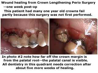
Wound healing following crown lengthening periodontal gum surgery. One week post op.
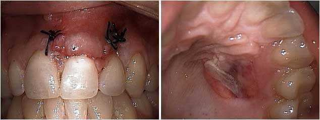
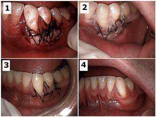
Wound healing three days after a free gingival gum graft. The upper front central incisor exhibited a lot of gum recession that resulted in a cosmetic defect when the patient smiled. 1) The gum graft is shown after being sutured in place. These black sutures stitches will be removed in about ten days. 2) The palate of the same patient showing the donor site for the graft. A gingival graft typically involves removing a small amount of palatal gingiva and sewing it where it is needed.
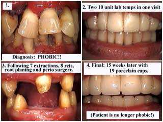
Full Mouth Reconstruction on a dental phobia patient. The third photo shows wound healing following 7 teeth extractions, 8 root canals and composite cores, full mouth root planing and then periodontal gum surgery and 19 units of bridgework (19 dental crowns) over 8 abutment teeth.
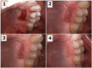
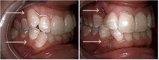
Free gingival graft of a lower left canine tooth. Wound healing of the palatal donor site. 1) Initial periodontal gum surgery site. 2) One week later. 3) Two weeks later. 4) Seven weeks later. Photo #1 of 2.
Free gingival graft of a lower left canine tooth. Wound healing of the recipient site. 1) Initial periodontal gum surgery site. 2) One week later. 3) Two weeks later. 4) Seven weeks later. Photo #2 of 2.
Bridge repair and periodontal wound healing. 1) Fitting the bridge over the bonded repair. 2) The bridge was cemented with composite resin. The mesial wing was also bonded to the distal of the lateral. 3) The gingivectomy was chosen over a flap because the patient had a low lip line and so the development of a pseudopocket with a flap was avoided. The red indicates where the gum line could have been. 4) The final result. The patient is informed of the need for bridge replacement when finances allow.