Before and after photos on how to prepare or drill porcelain laminates performed in our Porcelain Veneers office.
Teeth preparation – drilling – for ten lower porcelain veneers teeth laminates. Note that the upper ten dental veneers are already on.
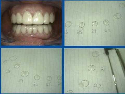
How to place and cement ten lower porcelain teeth veneers pictures. The upper ten porcelain dental veneers are already on. It is worthwhile to confirm the fit of each tooth veneer to the master dies and carefully separate them before placement in the mouth. It is very easy to confuse one with another and you only have about ten seconds before the dental bonding cement hardens!!
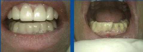
A simple method for how to make provisional temporary teeth veneers. You can use a clear acrylic stent that is relined with Luxatemp or you can just place the Luxatemp in the starting alginate impression that was made before teeth preparation. Luxatemp does not cure exothermically so it can be allowed to harden on the teeth without concern to the teeth nerves – root canal. The second photo shows half of the temporary teeth veneers removed.
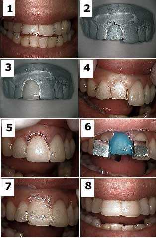
The “Bobbi” porcelain veneers method involves the fabrication of the teeth veneers on a master model BEFORE preparation of the actual teeth. This allows the tooth preparation and final porcelain dental veneers to be completed in a one-hour visit. 1) Pre-op view showing traumatic fracture – a broken tooth – of a porcelain veneer on tooth #8. 2) The tooth veneer preparation – drilling – is first made on the master model. 3) The porcelain dental veneer is fabricated on this master model. 4) Tooth preparation. 5) Initial seating of the porcelain veneer. 6) Etch with 37% phosphoric acid. 7) Vaseline placed on labial surface during seating to prevent dental bonding material from adhering to the labial surface of the porcelain. 8) Final result. The main drawback to this technique is having confidence that your preparation on the tooth will closely match that performed on the master model. Otherwise, the dentist will be faced with double lab charges if the teeth veneers do not fit. Note this technique can be performed only for porcelain teeth veneers where the expectation of marginal fit is not as high as for porcelain fused to metal dental crowns and this discrepancy is compensated for with the dental bonding system used for adhesion.
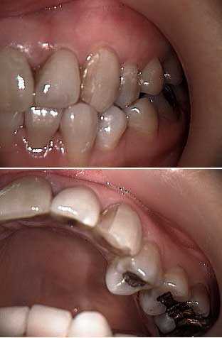
Porcelain laminate tooth veneer failure on an upper left canine. Notice the edge-to-edge occlusion bite in this area and that the original dental laminate did not wrap around to the palatal surface of the tooth. When correcting a cross bite with porcelain teeth laminates the dentist should create extra mechanical retention in the tooth preparation to help reduce sheer stresses on the dental bonding cement.
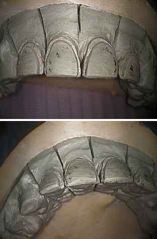
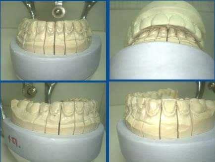
Four Maxillary Porcelain Teeth Veneers on a Master Model from the Dental Laboratory. The tooth
preparation – drilling – extends to the interproximal area to hide the cement line. The gingival margin – gum line – preparation needs to be very obvious to the technician. The teeth impression should extend into the gingival sulcus.
Try to create a positive seat in the preparation so the porcelain dental veneers won’t slip during cementation.
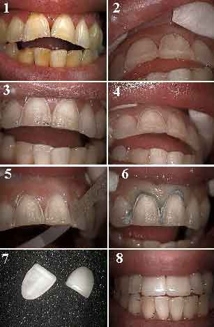
How to prepare – drill – two upper front teeth for porcelain teeth veneers. Before and after pictures.
1) Before photo with two broken central incisor teeth and an anterior open bite occlusion. Teeth whitening was already completed as was a dental prophylaxis. 2) Facial depth cut for the porcelain veneer on tooth #9. 3) Initial teeth preparation on #8 and #9. 4) Final teeth preparation on #8 and #9. 5) Slightly reducing interproximal contacts with sandpaper strip. 6) Retraction cord gently packed before taking a teeth impression. 7) The final porcelain dental veneers. 8) After picture with the porcelain teeth veneers cemented.
1) Before photo with two broken central incisor teeth and an anterior open bite occlusion. Teeth whitening was already completed as was a dental prophylaxis. 2) Facial depth cut for the porcelain veneer on tooth #9. 3) Initial teeth preparation on #8 and #9. 4) Final teeth preparation on #8 and #9. 5) Slightly reducing interproximal contacts with sandpaper strip. 6) Retraction cord gently packed before taking a teeth impression. 7) The final porcelain dental veneers. 8) After picture with the porcelain teeth veneers cemented.
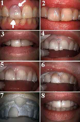
These pictures show how to make two upper front porcelain teeth veneers following an accident that broke these two teeth. Photo 1) Tooth #8 shows a burst blood vessel through enamel and deep cracks. Photo 2) Following root canal therapy and “walking” internal bleach. Photo 3) Following external office bleach – teeth whitening – of all teeth. Photo 4) Beginning incisal reduction for porcelain teeth veneers. This patient wanted to eliminate the facial dental bonding on tooth #9 and the deep crack lines. Photo 5) Beginning facial reduction using depth cuts. Photo 6) Final preparation for porcelain dental veneers. Photo 7) Porcelain veneers on the master model. Photo 8) Porcelain teeth veneers laminates cemented.