Before and after photos on study models diagnostic casts and master models performed in our TMJ Bite Guards office.
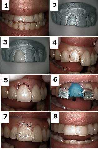
The “Bobbi” porcelain teeth veneer technique involves the fabrication of the dental veneer on a master model BEFORE preparation of the actual teeth. This allows the teeth preparation and final porcelain veneer to be completed in a one-hour visit. 1) Pre-op Before Photo shows a broken upper front tooth veneer #8. Photo 2) The veneer preparation drilling is made on the master models. 3) The veneer is fabricated on the master die from this master model in the dental laboratory by the porcelain technician. 4) Tooth preparation. 5) Initial seating of the porcelain veneer. 6) Etch with 37% phosphoric acid. 7) Vaseline placed on labial surface during seating to prevent bonding material from adhering to the labial surface. 8) After photo Final result. The main drawback to this technique is having confidence that your drilling preparation on the tooth will closely match that performed on the master models. Otherwise, the dentist will be faced with double lab charges if the veneer does not fit. Note this technique can be performed only for veneers where the expectation of marginal fit is not as high as for porcelain fused to metal dental crowns and this discrepancy can be compensated for with the bonding system used for adhesion.
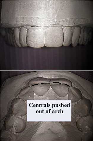
Study models dental casts show before and after orthodontics teeth braces treatment of a constricted narrow jaw in an adult female. Before Pictures. The narrow jaw makes the central teeth protrude labially – towards the lip. This makes the two front teeth overly prominent. The patient elected to treat this with braces. Photo #1 of 2.
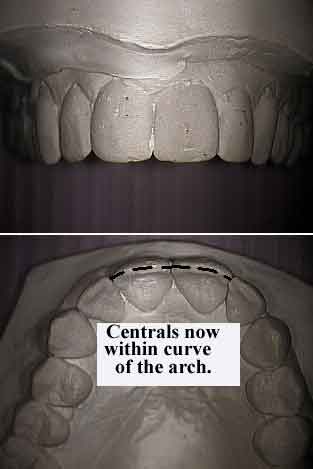
Study models dental casts show before and after orthodontics teeth braces treatment of a constricted narrow jaw in an adult female. After Pictures. The treatment time was six months. The upper front teeth were brought back into the “U” shape of the jaw that can be seen more easily in the bottom photo. This dramatically enhanced the patient’s smile. In a developing child it would be possible to provide even greater arch curvature than seen here in the plaster models. Photo #2 of 2.
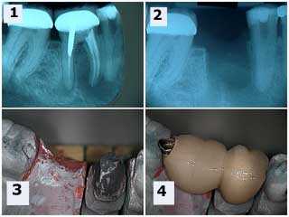
These x-rays and pictures show the fabrication of a two teeth bridge with the master working model and dies made from diestone. The occlusal rest seat preparation can be seen on the mesial of the second molar dental crown. This eliminated the need for removing the second molar crown. The premolar received root canal therapy following healing of the tooth extraction site. The patient did not want a dental implant.
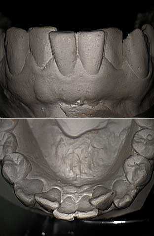
These photos show a frontal and occlusal view of a plaster study model of the lower arch or jaw. This diagnostic cast demonstrates lateral incisor teeth crowding in the mandible. These photos may also demonstrate post-orthodontic relapse associated with not wearing teeth retainers.
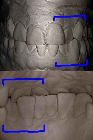
These photos show a frontal and lingual view of a plaster study model of the same dentition. Note the anterior cross bite between the maxillary left lateral and the mandibular left canine teeth. Sometimes a lot of information can be obtained by turning the diagnostic casts around and look from the inside out as if you were standing on the tongue. It is also a great teaching aid for patients.
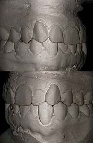
Lateral views of the diagnostic models of the same dentition. Note the anterior cross bite between the upper lateral incisor and the lower canine teeth on one side. A fixed orthodontic appliance – removable teeth braces – could be used to correct this cross bite.
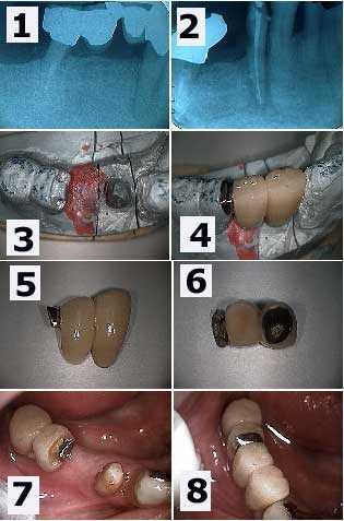
Cervical tooth decay and root canal infection around a lower right first premolar tooth. How to pictures and x-rays shown with a master working die stone model. 1) A large radiolucency is seen in the xray. Note the distal abutment is the distal root of a first molar following hemisection. 2) Following root canal treatment. The cervical dental caries can now be seen more easily with the teeth bridge removed. 3) The working master model shows the preparation of the mesial part of the distal two-unit dental bridge. This preparation will allow rigid connection to the new anterior bridge being made. 4) The new bridge section in place on the working model made of die stone. 5) & 6) Two photos of the new bridge showing the distal attachment. 7) An intraoral photo which mirrors the master model view seen in 3). 8) The final result. This patient preferred to see the gold occlusal rest. Otherwise, it could have been hidden under porcelain.
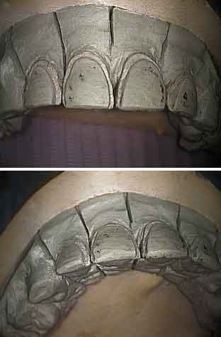
Teeth preparation photos for four upper porcelain dental veneers shown on a master working cast. The preparation – teeth drilling – extends to the interproximal area where the adjacent teeth touch to hide the dental cement line. The gingival margin preparation on the master model needs to be very obvious to the technician after the die stone model hardens and is removed from the impression. The impression should extend into the sulcus to help the lab tech identify both the final margin length and width. Try to create a positive seat in the prep so that porcelain teeth veneers won’t slip during dental cementation.
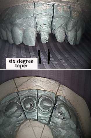
All porcelain crown preparation pictures for two upper central incisor teeth shown on a master model. The width of the shoulder should be around 2 mm. Over preparation will weaken the tooth; under preparation will weaken the porcelain. The dental impression should extend into the gingival sulcus to give the lab technician a clean finish line on the dies of the master cast.
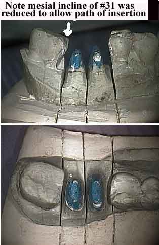
These photos show preparation -drilling – of tooth #30 following hemisection on a master die stone model. The individual tooth roots should be prepared for relative parallelism as seen in this working cast. The individual metal copings will join in a metal furcation. Porcelain will then be added to make the final dental crown look like one tooth.
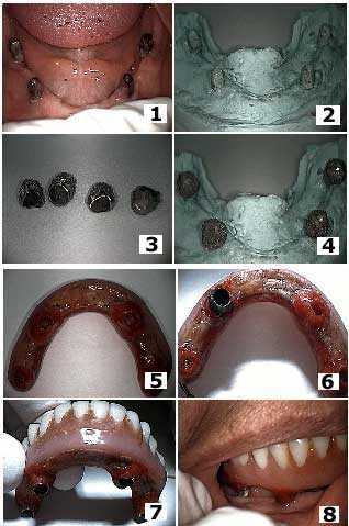
Retrofitting telescopic copings under an all-acrylic implant denture over a full mandibular subperiosteal dental implant. This implant and prosthesis is over 15 years old. How to pictures. The all-acrylic prosthesis had been successfully retained with a small amount of denture adhesive over the dental implant abutments. The patient would return about twice a year to reline the acrylic of the prosthesis that would wear over time against the dental implant posts. The patient wanted to reduce the frequency of these relines. 1) Photo of the full mandibular subperiosteal dental implant. 2) Photo of the master model die stone. 3) Photo of the four telescopic copings that were fabricated to fit over the implant posts and had roughened outer surfaces for incorporation into the acrylic prosthesis. 4) The four copings on the working model. 5) The underside of the all-acrylic implant prosthesis. 6) One coping being incorporated into the prosthesis at a time. The acrylic in the immediate area had been removed to allow the prosthesis to seat over the coping, then flowable self-curing acrylic was relined over it. 7) The prosthesis with all four copings attached. 8) An intraoral view of the dental implant prosthesis with the telescopic copings attached.
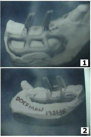
Photos of a unilateral maxillary subperiosteal dental implant on a master model. This master working model is made from a direct bone impression with soft tissue sutured out of the way. The patient will typically return in three weeks after custom fabrication of the implant.
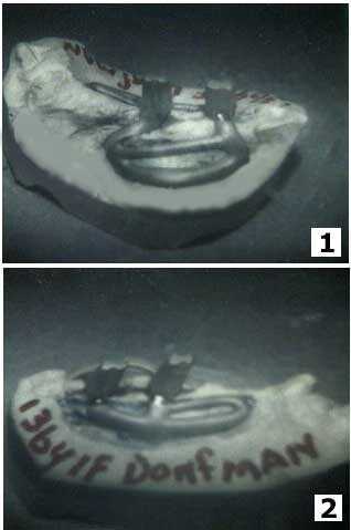
Pictures of a unilateral maxillary subperiosteal dental implant on a master working model. This type of impression requires the removal of a significant amount of periosteum so these dental models need to be treated with extreme care.
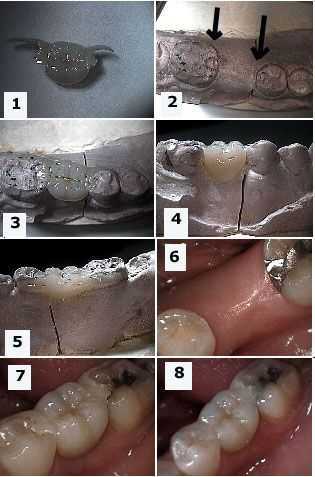
These master model pictures show how to fabricate a Maryland bridge in the mouth and on the master working model. 1) Photo of the Maryland bridge. 2) This master model photo shows the prepared teeth abutments with occlusal rests. 3) – 5) Photos of the dental bridge in place on the master cast. 6) Intra oral view of the edentulous span. 7) – 8) The bridge in place.