Before and after photos on combined root canal and gum problems performed in our Root Canal office.
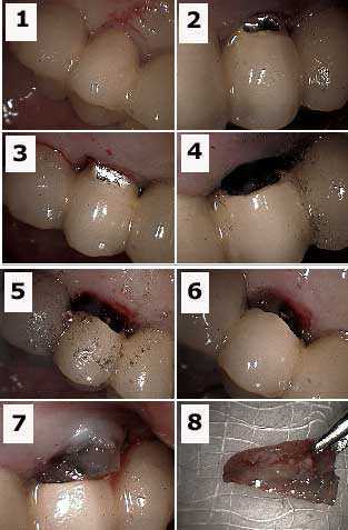
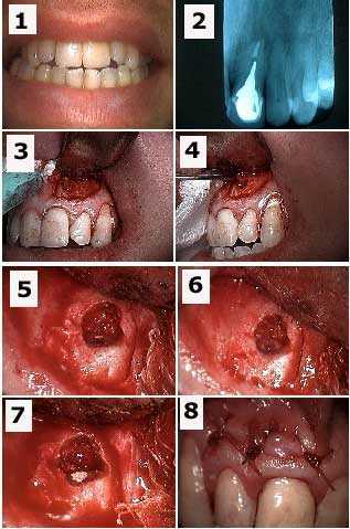
Endodontic periodontal lesions, combined root canal gum problems. How to pictures root resection from under a porcelain fused to metal dental bridge. 1) Labial photo of a tooth bridge abutment with an endo-perio lesion. 2) & 3) Initial preparation into porcelain teeth with a diamond bur. 4) Labial photo showing preparation. The metal portion of the bridge is prepared with a steel bur. 5) Palatal photo showing preparation. 6) Labial photo showing the tooth root under the porcelain (gutta percha is visible in the tooth root). 7) Tooth root being extracted. The occlusal height of this root must first be reduced to allow it to exit. 8) The extracted tooth root.
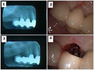
Endodontic periodontal lesions, combined root canal gum problems. Root resection from under a porcelain fused to metal dental bridge. 1) The x-ray shows the distal tooth abutment with a combined endodontic periodontal lesion. 2) Labial photo of the same tooth. 3) Radiograph following resection (the metal chad in the area of the extracted abutment was later removed). 4) Labial photo following extraction. This area could be filled in with dental bonding after healing. The occlusion on the distal cantilever was reduced. It opposed a lower full arch teeth bridge so that supraeruption was not a concern. Treatment options include: i) sectioning and removal of the distal cantilever, ii) dental implants, or iii) reevaluate over time with the patient informed to reduce function in this area.
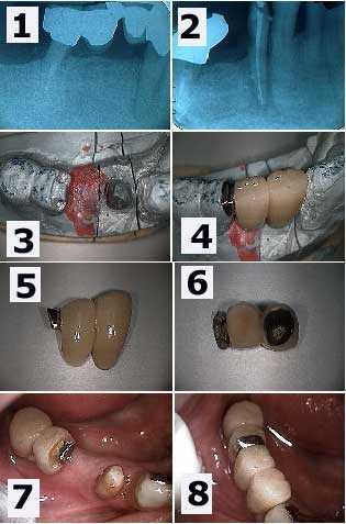
Endo perio lesions, combined root canal gum problems. Cervical tooth decay and root canal infection around a lower right first premolar. 1) A large radiolucency is seen in the x-ray. Note the distal tooth abutment is the distal tooth root of a first molar following hemisection. 2) Following root canal therapy. The cervical decay can now be seen more easily. 3) The working model showing the preparation of the mesial part of the distal two teeth dental bridge. This preparation will allow rigid connection to the new anterior teeth bridge being made. 4) The new dental bridge section in place on the working model. 5) & 6) Two pictures of the new bridge showing the distal attachment. 7) An intra oral image which mirrors the working model view seen in 3). 8) The final result. This patient was content to see the gold occlusal rest. Otherwise, it could have been hidden under porcelain.
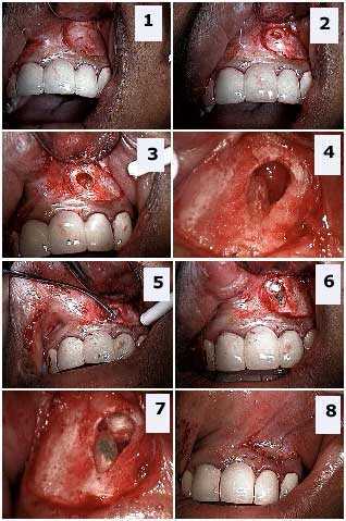
Endodontic periodontal lesions, combined root canal gum problems. Apicoectomy and retrograde tooth root apex filling. 1) Upper left lateral incisor tooth shown with tissue reflected. 2) Initial penetration through cortical bone. 3) Tooth apex located. 4) Tooth root after sectioning off the tooth apex. 5) Application of Mineral Trioxide Aggregate (MTA) retrograde filling material. 6) – 7) MTA in place. 8) Closure. The combined lesion completely healed in follow up examinations over six months.
Endodontic periodontal lesions, combined root canal gum problems. This tooth had a draining fistula – tooth abscess – and periodontal pocket around this upper front tooth. The fistula and pocket both healed after this procedure.