Before and after photos on direct pulp cap nerve exposure and pulpitis performed in our Root Canal office.
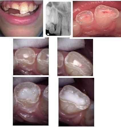
Direct pulp exposure of the dental nerve following traumatic injury in a child. Traumatic injury to teeth # 7 & 8. An Oral Surgeon ruled out other injuries. An Endodontist performed a partial pulpotomy (partial root canal therapy) on both teeth with MTA (middle photos). A fluoride-releasing glass ionomer was then placed on the remaining exposed dentin (lower photos). The intention is to achieve apexification. The teeth will then be prepared for full coverage crowns.
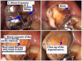
Direct exposure of the dental pulp tooth nerve in a broken silver filling that caused tooth decay. The tooth cavity caused tooth pain symptoms and an irreversible pulpitis. Exposure of the dental nerve was evident while removing the dental caries. Root canal therapy and a tooth crown is the most predictable dental treatment.
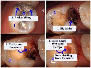
A broken silver amalgam tooth filling is removed using a dental drill and the pictures show dental caries into the tooth nerve dental pulp. This tooth needs root canal pulpitis treatment; bleeding is visible from the dental nerve. This patient had severe tooth pain symptoms.
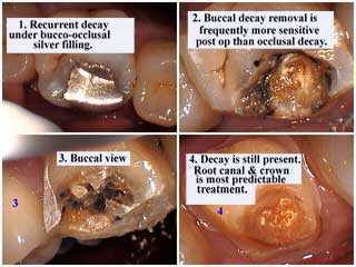
There is recurrent tooth decay under this silver filling. Note that buccal – cheek side – tooth decay removal with a dental drill usually causes more pulpitis symptoms afterwards than tooth cavity removal from the occlusal – biting – tooth surface.
Dental decay is still present in the pictures. Root canal treatment and placement of a dental crown is most predictable.
Dental decay is still present in the pictures. Root canal treatment and placement of a dental crown is most predictable.
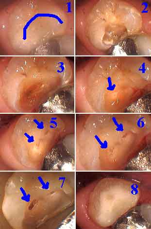
How to pictures. Direct pulp cap on a tooth nerve after direct exposure of the dental nerve while removing deep dental caries, a deep tooth cavity. This tooth needs root canal treatment and the placement of a tooth crown, dental cap.
1) Tooth decay is only seen in the x-ray.
2-7) Tooth cavity removal – tooth drilling pictures.
4) The tooth nerve exposure is first seen in this photo. 8) Dental bonding is completed as planned. More dental treatment is needed. This patient had an inflamed dental pulp and tooth pain symptoms. The pulp test showed the tooth was vital. It was agreed to first try to determine whether the tooth pain could be considered a reversible pulpitis that could heal with only tooth bonding or whether it would be considered an irreversible pulpitis needing root canal treatment. The depth of the tooth cavity suggested the latter and the patient agreed to return to meet with the Endodontist for a pulpectomy.