Photos and X-rays on endodontic retreatment by an endodontist or root canal specialist created in our Root Canal office.
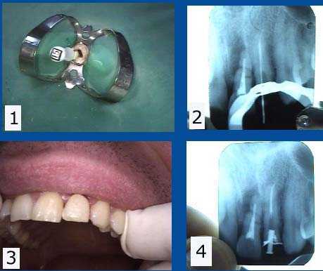
Endodontic root canal retreatment of an upper left lateral incisor tooth following fracture of an old dental crown. (The adjacent central incisor #9 will need to be extracted due to apical fracture). Photo 1) Tooth #10 – the upper left lateral tooth canal is widened with an endodontic file. Old gutta percha is removed using Chloroform solvent. A rubber dam clamp and rubber dam in place on the tooth to maintain sterility. 2) X-ray of an endodontic file marker used to measure vertical depth of the root canal to the tooth root apex. The prior gutta percha was not obturated – filled – to the root canal apex and the gutta percha had been left exposed to saliva after the dental crown fracture. Photo 3) Root canal filled with new gutta percha, temporary dental crown and temporary post re-inserted. 4) Post-op x-ray showing the obturated root canal and post space in preparation for a cast gold post and core impression.
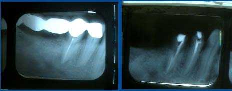
Endodontic root canal retreatment of tooth #29 and initial treatment of #28. This five teeth bridge had dental cement leakage and the teeth abutments developed cavities after only 1 and 1/2 years. The patient said that #29 was always sensitive after the original root canal was performed by her then general dentist. Root canal retreatment of #29 by an endodontist – root canal specialist – revealed a second, previously undiagnosed and untreated second root canal within the same tooth. A post space was created in this second canal after obturation.
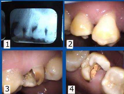
Tooth #13 has been left exposed for over a year since the root canal temporary filling fell out. 1. Current xray. 2. Buccal image. 3. Palatal image. 4. Occlusal image. Treatment options: Extraction and then tooth replacement or retreat the root canal, post and core, crown lengthening gum surgery and crown. This tooth is getting to the point where endodontic root canal retreatment is less favored and a dental implant may be more predictable.
Comparison of the before and after x-ray radiograph of endodontic root canal retreatment of a maxillary second premolar tooth. The root canal filling material did not reach the apex of the tooth root in the initial xray but now does in the final radiograph.
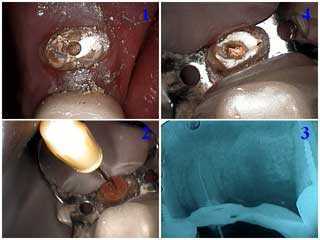
Root canal retreatment. Photo 1) Endodontic access is attained after removing old dental filling material. Photo 2) The root canal measurement file is placed. Photo 3) The measurement x-ray shows the file reaching the apex of the tooth root. This gives an accurate measurement of the canal length. 4) The xray shows the root canal is filled with gutta percha.
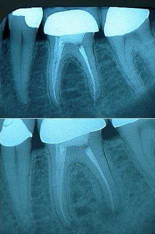
Before and after x-rays of a molar tooth after root canal endodontic retreatment. 1) The post op radiograph of the initial root canal looks good. A little root canal cement can be seen extruding from the tooth root apices. 2) A two year follow up xray shows the development of an expanding periapical radiolucency. This second x-ray shows the molar tooth after endodontic retreatment and the periapical radiolucency at the time of the retreatment.
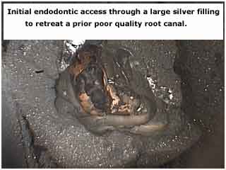
Endodontic root canal retreatment photo showing the initial endodontic access through a large silver dental filling.
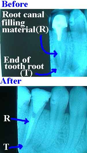
Before and After x-rays showing Root Canal Re-Treatment. The root canal filling material should extend to the end of the tooth root apex when properly performed. This is dental work better left to an endodontist, a root canal specialist.
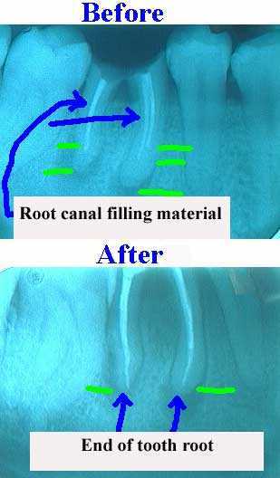
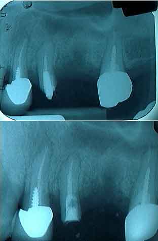
A Before and After xray showing Endodontic Root Canal Retreatment. Poor quality root canal therapy on the top does not show root canal filling material to the end of the tooth root apex. This allows bacterial leakage and infection. This is dental work better left to an endodontist, a root canal specialist.