Before and after photos on endodontics is needed for root canal pain infection performed in our Root Canal office.
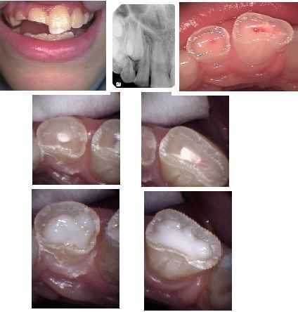
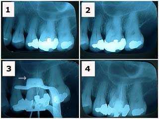
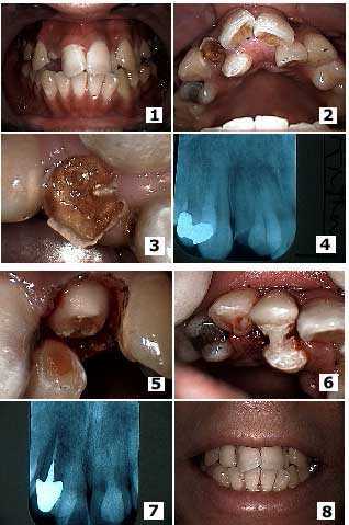
Pediatric Endodontics, Root Canal Therapy for children: Photos & x-ray. Traumatic injury to teeth # 7 & 8. Oral Surgeon ruled out other injuries. An Endodontist performed a partial pulpotomy on both teeth with MTA (middle photos) in order to facilitate closure of the root apices. A fluoride-releasing glass ionomer was then placed on the remaining exposed dentin (lower photos). The teeth will then be prepared for full coverage crowns.
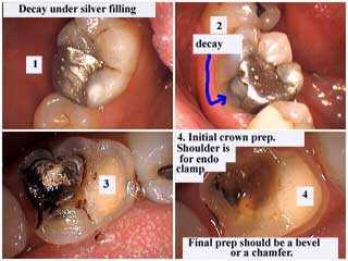
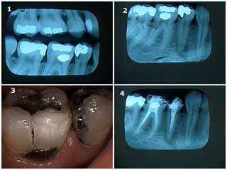
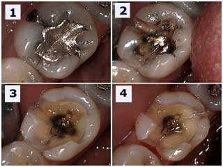
How to photos explain why root canal therapy (endodontics) is sometimes needed after removal of deep tooth decay (dental caries). The white material (in the top right picture) is a sedative medicine that was previously placed and left under the old silver filling to help reduce post operative sensitivity or pain. This was needed because the dentist’s drilling approached the nerve (root canal). When this medicine and the remaining cavity are removed (bottom left picture) the drilling is even closer to the nerve (root canal). Sometimes post operative sensitivity requires root canal therapy to treat the tooth pain. Root canal therapy should be performed by an Endodontist.
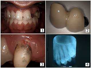
Need for endodontics root canal. A female patient presented with a dark upper front tooth. An endodontist performed root canal therapy and then cosmetic teeth whitening and internal walking bleaching performed to whiten the tooth.
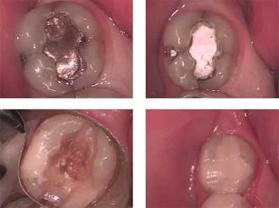
Need for endodontics root canal. This molar had a lot of tooth decay under an old amalgam silver dental filling. The endodontist pulp tested it – before any dentistry was performed – and found it was nonvital. The patient had experienced severe tooth pain and jaw pain – an acute root canal pulpitis.
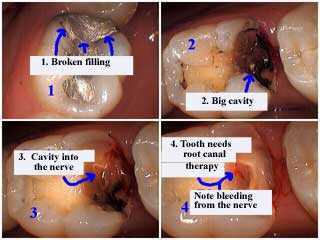
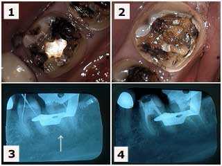
Need for endodontics root canal due to pulp nerve exposure and an irreversible pulpitis. A broken silver filling is removed showing a big tooth cavity into the nerve. This tooth needs root canal therapy; bleeding is visible from the nerve.
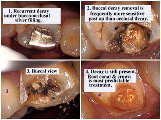
Need for endodontics root canal. There is recurrent tooth decay under the silver dental filling. Note that buccal decay removal is frequently more sensitive post-op than occlusal decay. Decay is still present. Root canal and dental crown is most predictable.
This tooth needed endodontics root canal because of tooth decay that went into the nerve or pulp of the tooth.
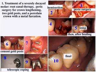
Treatment of a severely decayed molar:
– root canal therapy.
– gum surgery for crown lengthening.
– two cast gold post and cores.
– porcelain dental crown with a metal furcation.
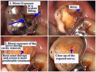
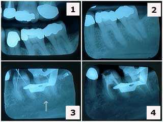
Direct exposure of the root canal under a broken silver filling caused by tooth decay. Exposure of the tooth nerve was evident while removing the decay. Root canal therapy and dental crown is the most predictable treatment.
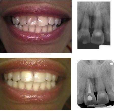
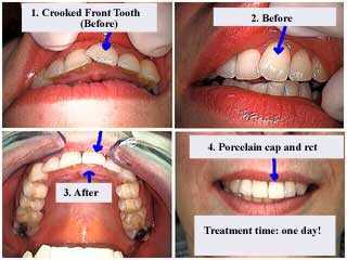
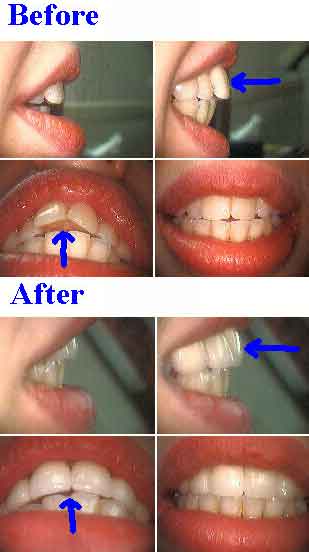
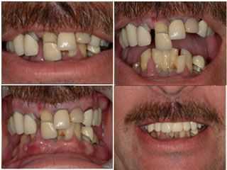
Cosmetic dentistry can sometimes require endodontics root canal when a patient declines teeth braces. This patient wanted to treat the crooked front tooth seen in the top two pictures without teeth braces. The tooth was brough back into alignment after root canal and a porcelain tooth cap as seen in the bottom photos. Treatment time could be 24 hours.
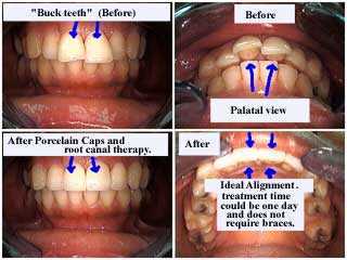
Top two central teeth were brought back into alignment with porcelain dental crowns after root canal.
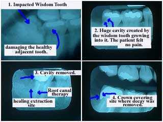
Need for endodontics root canal caused by an impacted wisdom tooth growing into the adjacent tooth and causing dental caries. The patient did not feel pain.
After the wisdom tooth was extracted the second molar received root canal and a dental crown (the crown covers the site where tooth decay was removed.
There is tooth decay under this silver tooth filling that went into the tooth nerve, a direct pulp exposure that resulted in an irreversible pulpitis. This tooth needs endodontics root canal therapy.
Direct exposure of the nerve while dental bonding a tooth with a large and deep cavity. This tooth needs root canal therapy and a dental crown.
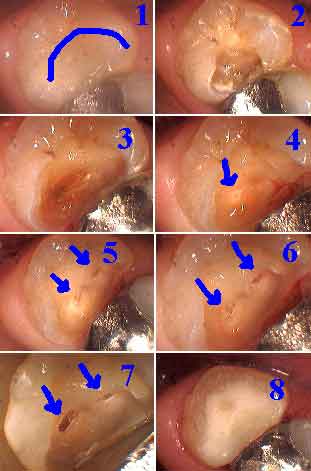
1) Tooth cavity only seen in this photo.
2-7) Cavity removal by drilling tooth.
4) Tooth pulp nerve direct exposure first seen.
8) Dental bonding completed. This patient was informed of the recommendation for root canal but given the option when to proceed by completing the dental bonding as planned.
Need for endodontics root canal – cosmetic dentistry. This patient had allergic dermatitis and could not tolerate teeth braces. She wanted to retract her upper front teeth. Aesthetic dentistry included vital root canal and porcelain teeth crowns on these teeth.
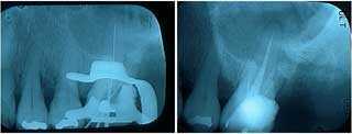
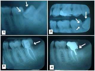
Need for endodontics root canal – this patient had a tooth abscess, irreversible pulpitis and swollen jaw. X-rays of root canal therapy showing initial lengths and then final obturation in an upper left first molar tooth.
Severe tooth decay causing tooth pain and a draining tooth infection that needs endodontics root canal.
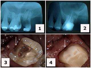
Treatment of a severely decayed upper left first molar. 1) Initial x-ray. 2) X-ray following extraction of tooth #15 and root canal therapy on tooth #14. Note the gingival extent of the tooth decay. 3) Tooth preparation following root canal therapy and the removal of the distal decay. 4) Final tooth preparation of the composite crown buildup. Note how the distal tooth preparation extends gingivally beyond the composite to tooth.
1) & 2) X-rays of an upper left molar tooth that needs endodontics root canal shows a large silver dental filling in a patient who had been experiencing tooth pain, infection and a swollen jaw. Note the decreased size of the pulp chamber in this tooth. 3) X-ray of endodontic file lengths. Note that the calcification in the mesial root of #14 initially prevented instrumentation. 4) Final endodontic obturation showing that the mesial root of #14 was located and treated. Chronic inflammation in the dental pulp due to the presence of large old dental fillings can increase the difficulty of root canal therapy. Earlier root canal therapy should be considered in these situations.
Need for endodontics root canal. Emergency teeth pain infection and a swollen jaw in the lower right quadrant. 1) X-rays show big teeth cavities in the second premolar and first molar and smaller cavities in both the second and third molars. 2) There was also a large periapical radiolucency around the root apex of the second premolar. 3) Photo of the second premolar and first molar. 4) Radiograph of the second premolar and first molar following root canal therapy. The patient felt better immediately following this. Note: these teeth were treated following a careful differential diagnosis. It would not be surprising, if in another patient, the pain came from a tooth with a less apparently severe problem.
Need for endodontics root canal due to an irreversible pulpitis. 1) & 2) Two lower left molar teeth following removal of large, old silver dental fillings in a patient who had been experiencing teeth pain infection and abscess. 3) X-ray of endodontic file lengths. Note that the calcification in the mesial root of #18 initially prevented instrumentation. 4) Root canal obturation shows that the mesial root of #18 was located and treated. Chronic inflammation in the dental pulp due to the presence of large, old fillings can increase the difficulty of root canal therapy. Earlier root canal therapy should be considered in these situations.
Tooth decay and tooth pain – need for endodontics root canal. 1) & 2) X-rays of tooth #’s 19 & 18 showing large amalgam fillings. This emergency patient had a lot of teeth pain. Note the decreased size of the pulp chambers in these teeth. 3) X-ray of endodontic file lengths. Note that the calcification in the mesial root of #18 initially prevented instrumentation. 4) Final endodontic obturation showing that the mesial root of #18 was located and treated. Chronic inflammation in the dental pulp due to the presence of large, old fillings can increase the difficulty of root canal therapy. Earlier root canal therapy should be considered in these situations.
Need for endodontics root canal – tooth decay removal under a large amalgam silver filling revealed a fracture line. Photo 1) The large silver filling showing a mesial interproximal crack line. Photo 2) Amalgam removal still shows the presence of the crack line. Photo 3) This mesial crack line is now seen to be continuous through the middle of the tooth and out towards the buccal. Photo 4) The crack line has been removed as much as possible without invading the pulp chamber, the tooth nerve. Root canal therapy and a dental crown would be the most predictable method to keep the mesio-buccal cusp from further fracture. Tooth dental bonding would be a second choice that would help to prevent further cracking particularly if the mesio-buccal cusp was reduced occlusally and bonding overlayed. A non-bonded amalgam silver filling would probably exacerbate the fracture over time.
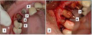
Treatment of a broken lateral incisor with tooth decay that needs endodontics root canal adjacent to a palatally-displaced supernumerary tooth. 1) – 2) Initial presentation. Note dental caries on the lateral tooth and the palatal location of the adjacent supernumerary tooth. 3) Close up of the broken, tooth cavity lateral and the adjacent palatally-displaced supernumerary tooth. 4) Radiograph. 5) The same tooth after gum surgery and initial tooth preparation. 6) Placement of the temporary cap. Note that acrylic was extended from the temporary on the lateral to the supernumerary to provide initial stability of the temporary cap until root canal therapy and a cast post and core was placed. 7) Radiograph of the final root canal therapy, cast post and core and dental crown. 8) Final result.
Need for endodontics root canal – dental bridge repair and tooth decay into the pulp nerve. 1) – 2) Patient had a two tooth dental bridge on an upper canine abutment tooth, lateral incisor pontic and palatal wing attached to the distal of the central incisor tooth. The patient presented with the teeth bridge out. 3) Initially it seemed that there was external resorption on the facial surface of the canine near the gingival margin. 4) Initial x-ray.
An irreversible pulpitis needs endodontics root canal because of tooth decay caused by an overretained wisdom tooth growing into a second molar damage.
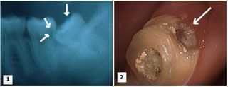
1) – 2) Two x-rays showing a lower wisdom tooth impacted on an angle and pushing into the adjacent second molar causing a large cavity. 3) X-ray shows the wisdom tooth was removed and the second molar following root canal therapy with the large distal cavity still present. 4) Cast gold post and core and crown in place. Note the distal dental crown margin completely covers where the distal tooth cavity was removed.
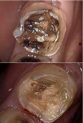
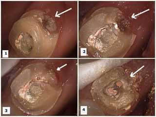
These pictures show tooth drilling of the second molar to remove the distal tooth cavity after root canal endodontics was performed on it and the wisdom tooth was extracted. Photos 1) – 2) Dental caries is present in the distal area of the second molar. Temporary filling material is visible in the occlusal opening following root canal therapy. 3) Dental decay is removed. 4) Temporary filling material removed showing gutta percha in root canal orifices prior to cast post and core preparation.
Need for endodontics root canal – a huge dental cavity caused by a wisdom tooth growing on an angle into it. 1) X-ray showing a lower wisdom tooth impacted on an angle and pushing into the adjacent second molar causing a large tooth cavity. 2) Tooth decay is present in the distal area of the second molar. Temporary filling material is visible in the occlusal opening following root canal therapy.
Oral reconstruction of this patient will certainly include endodontics root canal therapy to treat the infection and teeth abscesses noted during the exam. The patient did not feel teeth pain!
Need for endodontics root canal. Selective extraction during an upper reconstruction. The initial treatment plan considered saving #’s 6, 7 and 9 while extracting #’s 5 and 8. During the surgery, #5 was considered more stable than #6 so #5 was retained and #6 was extracted. The retained teeth will be used to support a fixed temporary bridge until subsequent implant dentistry is completed.