Our endodontist team includes 16 cosmetic dentists, specialists including two endodontists – root canal specialists – and lab ceramists. In particular our low-volume office focuses on high-end care. Therefore our treatment is quick and comfortable. In addition our MD-anesthesiologist offers several options for dental sedation and nitrous oxide laughing gas. We offer intelligent and honest diagnosis based upon 31+ years of experience. Photos and X-rays on how our endodontist properly performs root canal therapy was created in our Root Canal office.
endodontist teaches how to properly perform root canal therapy
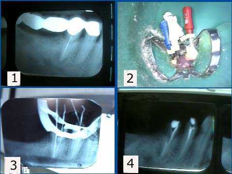
In endodontics obturation means the root canal filling procedure. 1. This x-ray shows periapical pathology – root canal infection – in tooth #29. Patient had chronic tooth pain following her original root canal therapy. Photo 2. A second root canal was found with endodontic files. 3. This x-ray shows an endodontic file in the second root canal. 4. Post preparation in the second canal of #29 after endodontic obturation with gutta percha and lateral condensation.
endodontist teaches how to properly perform root canal therapy
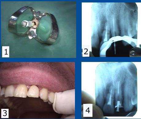
In endodontics obturation means the root canal filling procedure. Root canal retreatment of an upper left lateral incisor tooth following a broken tooth and fracture of an old dental crown. The adjacent central incisor #9 will need to be extracted due to root apex fracture. 1) Tooth #10 – the upper left lateral incisor tooth root canal was widened with an endodontic file. Chloroform solvent helps to remove old gutta percha. A rubber dam clamp and rubber dam are in place on the tooth to maintain sterility.
2) Radiograph of an endodontic file marker used to measure vertical depth of the root canal to the root apex. The prior gutta percha was not obturated to the canal apex and the gutta percha had been left exposed to the oral saliva after the tooth crown fracture. 3) Root canal filled with new gutta percha and lateral condensation, temporary dental crown and temporary post re-inserted. 4) Post-op xray shows the root canal obturation and post space in preparation for a cast gold post and core impression.
Obturation endodontics root canal filling using gutta percha and mineral trioxide aggregate in the treatment of internal resorption
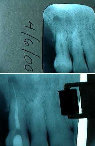
Internal resorption treatment for tooth #10. 1) Pre-op xray 11/15/2000. Patient had a history of falling off a horse. 2) Post-op x-ray 2/22/2001. #40 file used with NAOCL but #30 file used to apex. MTA mineral trioxide aggregate placed in the oval shaped void in the middle of the root canal. 3) xray Reevaluation 7/7/2004. The internal resorption appears stable.
endodontist explains the importance of the final root canal filling i.e. obturation
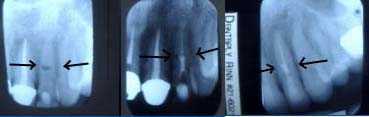
Obturation endodontics root canal filling. Before and after x-rays of a retreated molar tooth root canal. 1) The post op radiograph of the initial root canal looks good. A little root canal cement can be seen extruding from the tooth root apices. 2) A two year follow up x-ray showed the development of an expanding periapical radiolucency – periapical pathology – associated with tooth pain. The second xray shows the retreated molar and the periapical radiolucency at the time of the retreatment. This retreatment involved cleaning out all root canals to their apices and placement of new gutta percha root canal filling material with lateral condensation.
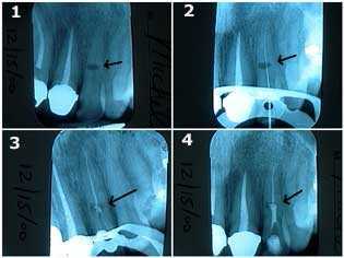
Root canal therapy involving internal resorption following an accident
Obturation endodontics root canal filling during treatment of internal resorption in a lateral incisor tooth following an accident. How to x-rays. 1) The oval-shaped radiolucency in the middle of the tooth length is the site of the internal resorption. The patient was informed of the guarded long-term prognosis of the tooth. 2) & 3) Traditional root canal therapy was first performed. The root canal space was cleaned and shaped, sterilized with NaOCL, and then filled with gutta percha – obturated – up to a point apical to the internal resorption. 4) MTA, Mineral Trioxide Aggregate, was then placed into the area of the internal resorption and coronal to it. Keeping the MTA moist will give more working time for condensation of material.
How to perform endodontics
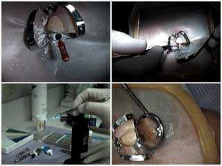
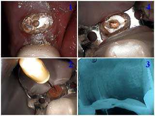
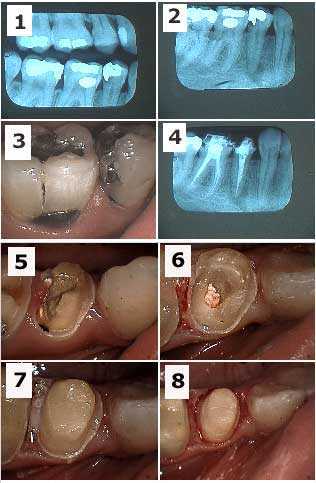
How to pictures show endodontic obturation technique in a maxillary canine tooth. 1) The endodontic file is in place within the root canal prior to the measurement x-ray radiograph. 2) Lateral condensation – root canal filling – after the gutta percha master cone is placed with root canal cement. 3) and 4) Heating the endodontic condenser to vertically condense and sever the gutta percha at the pulpal chamber floor.
endodontist explains the importance of the final root canal filling i.e. obturation
Root canal obturation i.e. filling is one measure of the quality of the procedure
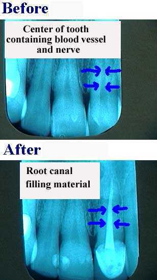
Understanding Root Canal Therapy quality by seeing the extent of Obturation Endodontics root canal filling in a dental x-ray. The root canal filling material should extend to the very end of the tooth root when endodontics is properly performed.
endodontist explains the importance of endodontic access
endodontist explains root canal retreatment i.e. fixing a previous bad root canal
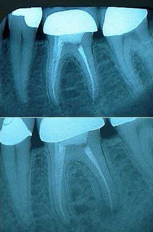
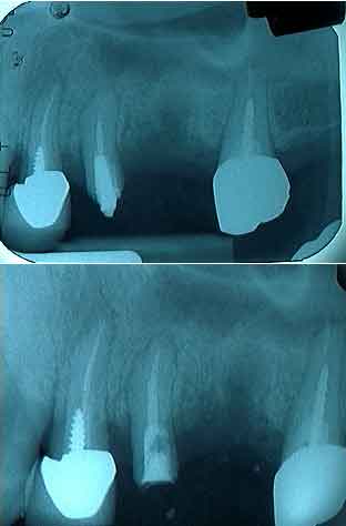
Comparison of the initial and final radiograph of root canal retreatment of a maxillary second premolar tooth. The root canal filling material does not reach the apex of the tooth root in the initial x-ray but now does in the final radiograph. Dental patients should ask to see the post op x-ray after root canal therapy because the obturation endodontics root canal filling should be relatively easy to judge for a non-dentist.
endodontist explains root canal retreatment i.e. fixing a previous bad root canal
endodontist show how root canal therapy is performed through an existing crown
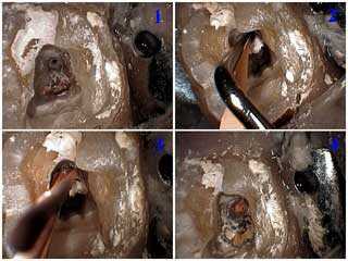
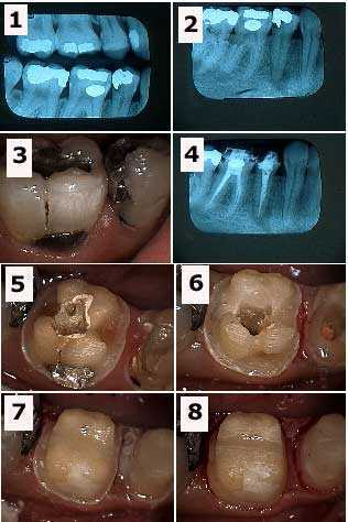
How to achieve endodontics root canal access through a dental crown on an upper right lateral incisor tooth. Pictures. 1) Drilling through the crown provides endodontic access. 2) & 3) Placement of the primary gutta percha point. 4) & 5) Lateral condensation of the accessory gutta percha points – the root canal filling material. 6) & 7) Heating the endodontic condenser to vertically condense and sever the gutta percha at the pulpal chamber floor. This completes endodontic obturation. 8) Placement of the temporary tooth filling.
Root canal tooth pain treatment
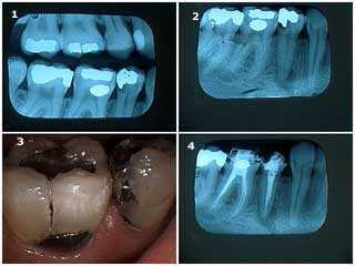
Emergency teeth pain in the mandible. 1) Radiographs showing big teeth cavities in the second premolar and first molar and smaller teeth cavities in both the second and third molars. 2) There was also a large periapical radiolucency around the apex of the second premolar. 3) Photo of the second premolar and first molar. 4) Xray radiograph of the second premolar and first molar following root canal obturation. The patient felt better immediately following root canal. Note: these teeth were treated following a careful differential diagnosis by the Endodontist. It would not be surprising in another patient if the tooth pain came from a tooth with a less apparently severe problem.
endodontist explains root canal tooth pain treatment
endodontist explains the importance of the final root canal filling i.e. obturation
Root canal under crown and bridge repair
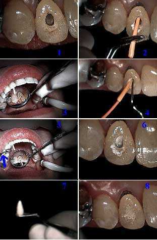
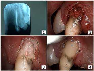
Teeth Bridge repair pictures with a post-op xray showing good obturation endodontics root canal filling. This patient wanted to save his teeth bridge until he got another job . 1) X-ray after root canal therapy shows nice endodontic obturation from the Endodontist. 2) Gingivectomy with electrosurgery. Note the still unusual look of the facial tooth decay. 3) Following tooth decay removal. Note the communication to the gutta percha root canal filling material. 4) The dental bonded restoration.
In conclusion Dr. Jeffrey Dorfman created all of the dentistry shown on this 4,400 page website. In brief we offer intelligent & honest diagnosis and better results for people seeking a endodontist. Visit us when you want it done right the first time; you will save money by initially spending more. Therefore please call The Center for Special Dentistry®.