Our root canal tooth pain treatment team includes 16 cosmetic dentists, specialists (including two endodontists) – root canal specialists – and lab ceramists. In particular we are a low-volume office focused on high-end care. Therefore our treatment is quick and comfortable. In addition our MD-anesthesiologist offers several options for dental sedation and nitrous oxide laughing gas. We offer intelligent and honest diagnosis based upon 31+ years of experience. Photos and X-rays on root canal tooth pain treatment – endodontics created in our Root Canal office.
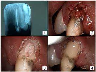
Root canal tooth pain treatment for an irreversible pulpitis i.e. root canal infection
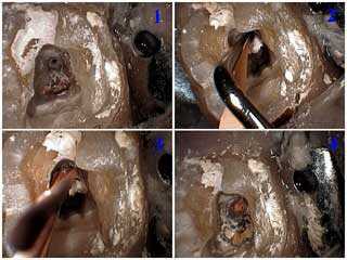
Endodontics shows treatment for an irreversible pulpitis. 1) Endodontic access attained and showing the distal and two mesial root canals. 2) Next lateral condensation of gutta percha in the distal canal after the master cone was cemented in place. 3) Then the auxiliary point is placed in the distal canal after the spreader was removed. 4) Lastly all three canals are obturated with gutta percha. A cotton pellet and Cavit will be placed as a temporary dental filling.
Root canal tooth pain treatment instrumentation
Root canal tooth pain treatment technique
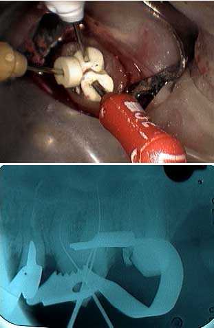
Endodontic technique in an upper left canine tooth. Photo 1) The endo file is in place within the root canal prior to the measurement radiograph, x-ray. Photo 2) Lateral condensation of gutta percha occurs after placement of the master cone with root canal cement.. Photos 3) and 4) Heating the endodontic condenser to vertically condense and sever the gutta percha at the pulpal chamber floor.
Root canal tooth pain treatment technique
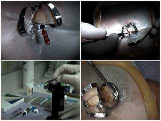
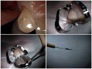
1) How to endodontic technique in an upper left canine tooth. 2) Surround the tooth with a rubber dam and a butterfly clamp. 3) This mirror image shows the obturated root canal. 4) The extirpated pulpal tissue is shown.
A before and after X-ray example of good root canal tooth pain treatment
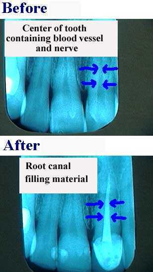
Another before and after X-ray example of good root canal tooth pain treatment
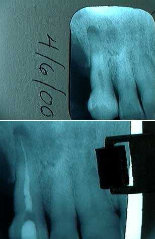
In brief Endodontics focuses on the dental specialty of tooth pain treatment. Extend the root canal filling material or gutta percha to the end of the tooth root apex. This is proper endodontic therapy.
A pulpotomy during root canal tooth pain treatment
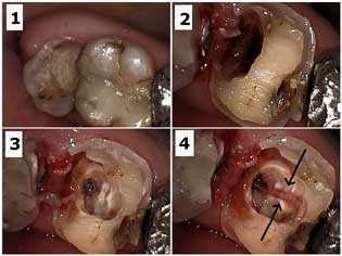
Endodontics, root canal therapy for irreversible pulpitis symptoms. Pulpotomy in an upper right first molar tooth. Photo 1) Initial image of the molar. Huge dental caries – tooth cavity – was visible in the x-ray. Photo 2) Initial preparation for tooth decay removal. Photo 3) Pulpotomy performed within the pulp chamber. Photo 4) Pulpal nerve tissue being removed (arrows).
Placement of a temporary filling after root canal tooth pain treatment
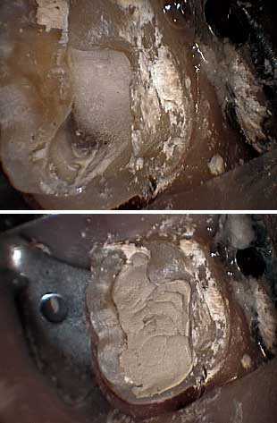
Endodontics for tooth pain. Placing cotton in the root canal access. This facilitates the location of the root canals during the subsequent restorative dental filling procedures. Placement of the temporary tooth filling.
An apicoectomy is another type of root canal tooth pain treatment that involves oral surgery
Preventive endodontics in root canal tooth pain treatment
Endodontics is the tooth pain treatment for dental pulpits, irreversible inflammation of the dental pulp
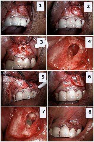
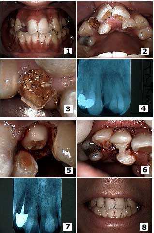
These pictures show treatment tooth decay in a broken upper lateral incisor tooth adjacent to a palatally-displaced supernumerary tooth. Photos 1) – 2) Initial presentation. Note the broken, decayed lateral and the palatal location of the adjacent supernumerary tooth. Photos 3) Close up of tooth decay in the broken lateral tooth and the adjacent palatally-displaced supernumerary tooth. 4) X-ray, radiograph. Photo 5) The same tooth after gum surgery and initial tooth preparation. Photo 6) Placement of the temporary dental crown. Note that acrylic extended from the temporary on the lateral to the supernumerary to provide initial stability of the temporary crown until root canal and a cast post and core was placed. Photo 7) Radiograph of the final root canal therapy, cast post and core and crown. Photo 8) Finally the aesthetic result the patient was seeking.
Always place a final restoration after root canal tooth pain treatment
Neglect of an endodontically treated molar tooth means that the tooth had been treated with root canal but the necessary additional dentistry like a dental crown was not done afterwards. Patients should understand that root canal is typically only one of several procedures that a tooth will need and not completing ALL procedures will likely lead to a premature and expensive failure that could result in tooth extraction.
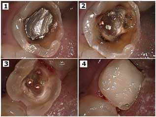
Treatment of a big tooth cavity in a molar that had previous root canal therapy. Dental bonding was being placed at this time – instead of a dental crown – until after the completion of teeth braces. 1) Neglected lower molar that had an adequate root canal yet large dental caries under the amalgam silver dental filling. 2) Photo of the extent of the tooth decay after removal of the silver filling. 3) Tooth decay removed. Note the gutta percha coming out of the three orifices. 4) The teeth bonding filling restoration. The point here is that a bonded restoration can be placed even when a crown is obviously indicated. Placement of the dental crown will occur after the patient completes orthodontics.
Complications with root canal therapy i.e. endodontics
Dental bridge repair showing tooth decay extending to the root canal filling material, gutta percha. 1) X-ray after endodontics. The root canal itself did not seem subject to external resorption. 2) Then gingivectomy. Note the still unusual look of the facial tooth decay. 3) Following dental decay removal. Note the communication to the gutta percha. 4) Lastly the teeth bonding dental restoration.
In conclusion Dr. Jeffrey Dorfman created all of the dentistry shown on this 4,400 page website. In brief we offer intelligent & honest diagnosis and better results for our root canal tooth pain treatment patients. Visit us when you want it done right the first time; you will save money by initially spending more. Therefore please call The Center for Special Dentistry®.