Photos and X-rays on symptoms diagnosis and treatment of internal resorption created in our Root Canal office.

Internal resorption treatment for tooth #10. The patient did not feel tooth pain symptoms because the nerve was non-vital. 1) Pre-op x-ray taken on Nov 15 2000. This patient had an accident falling off a horse. 2) Post-op x-ray taken Feb 22 2001. The Endodontist used a #40 file with NAOCL but a #30 file was used to the tooth root apex. MTA mineral trioxide aggregate was then placed. 3) Xray reevaluation July 7 2004. The internal resorption appears stable. Before and after treatment xrays of internal resorption. Note that internal resorption occurs from the inside of the tooth and spreads outward while external resorption occurs on the outside of the tooth and spreads inward.

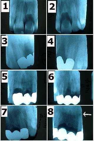
An unusual case of internal resorption occurring within several teeth of a 30 year-old male over ten years. 1) & 2) The patient related a history of trauma to the upper anterior teeth when these x-rays were taken in January 1991. Tooth #9 is undergoing severe internal resorption. 3) & 4) X-rays of the metal framework of a three teeth bridge in March 1991 following tooth extraction of tooth #9. 5) In May 1993 tooth #8 began showing symptoms of severe internal resorption. 6) & 7) X-rays of the metal framework of a four teeth bridge in July 1993 following tooth extraction of tooth #8. 8) Xrays of the teeth abutments #7 & 10 in April 2000 show a periapical radiolucency appearing around tooth #10. The patient was informed of the need for root canal therapy. X-rays in May 1995 & December 1997 had not showed pathologic changes.

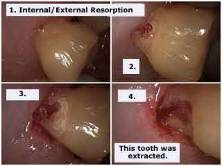
Internal and External Resorption in a maxillary molar tooth. Internal resorption occurs from the inside of the tooth and spreads outward while external resorption occurs on the outside of the tooth and spreads inward. These pictures show External Resorption. The diagnosis of Internal Resorption was made with a dental radiograph x-ray. This patient did not feel tooth pain symptoms.

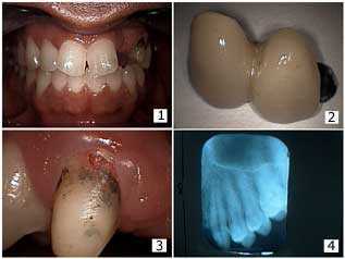
Dental Bridge repair and apparent External Resorption. This patient lost his job and could not afford a replacement bridge at this time. Pictures 1) – 2) Patient had a two-unit bridge on an upper canine abutment, lateral incisor pontic and palatal wing attached to the distal of the central incisor tooth. The patient presented with the bridge out and did not feel any tooth pain symptoms. 3) In this photo it appears that there was external resorption on the facial surface of the canine tooth near the gingival margin. 4) The initial x-ray did not show internal resorption. The Endodontist subsequently determined this was pulpal tissue and it was removed during pulpal extirpation. It was not external resorption.