Photos and X-rays on the tooth nerve is called the dental pulp created in our Root Canal office.

Endodontist treatment of internal resorption in a maxillary upper lateral incisor tooth #10. How to x-ray series. 1) Pre-op 11/15/2000. Patient had a history of falling off a horse. 2) Post-op 2/22/2001. #40 file used with NAOCL but #30 file used to apex. MTA – mineral trioxide aggregate – placed. 3) Reevaluation 7/7/2004. The internal resorption appears stable.

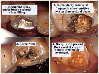
There is recurrent tooth decay under this amalgam silver dental filling. Buccal tooth decay removal – using a dental drill – frequently results in more post-op sensitivity or tooth pain than tooth decay removal from the occlusal – or biting – surface. This tooth pain may result in a reversible pulpitis that will heal without further dental treatment or it may result in an irreversible pulpitis and need root canal. Tooth decay is still present in this photo. Root canal and a tooth crown is the most predictable dental treatment to offer a patient in this example when drilling is close to the tooth nerve. A pulpotomy involves the removal of part of the dental pulp – to treat emergency tooth pain for example – but when there is not enough time to complete a full pulpectomy.