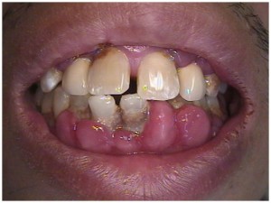
Treatment plans for Drug-induced gingival hyperplasia (DIGH)
February 23, 2010 4:09 pmMy exercise today was to look over a patients record and figure out what the possible treatment options are for the next phase of dentistry.
Chief Complaint: Patient wants to explore treatment options regarding possibility of restoration of anterior dentition. He is open to dentures, implants and any other options. Patient is very phobic, repeated past history of failed dentistry. No dental visits in the past several years.
Relative medical history: Patient has HTN and is taking a calcium channel blocker
Drug-induced gingival hyperplasia (DIGH):
Inflammation of the gingival tissue from bacterial plaque and the subsequent development of gingival crevicular fluid may allow sequestration of the calcium channel blocker, thus predisposing the tissue to a localized toxic effect and the development of gingival hyperplasia. All of the available calcium channel blockers have been reported to cause gingival hyperplasia.
Treatment options include meticulous plaque control, and in severe cases, gingivectomy. Drug-induced gingival hyperplasia (DIGH) is an iatrogenic dental disorder that is characterized by gums that are enlarged and inflamed, and bleed readily upon probing. The gums appear lobulated from papillary enlargement, and the tooth crowns may be partially covered by hyperplastic tissue. Drug-induced gingival hyperplasia is usually only cosmetically disfiguring; however, the formation of tissue pockets can interfere with proper oral hygiene, contributing to periodontal disease and dental caries. Patients who develop DIGH are at risk of treatment failure because of noncompliance. Those who develop severe DIGH may eventually require invasive oral surgery, such as a gingivectomy. (D.B. Lawrence et alJ Fam Pract 1994; 39:483-488)
Initial therapy consisted of scaling and root planing, extraction of four lower incisors that had severe bone loss, and provisional restorations in the edentulous space. After scaling and root planing was completed, and four lower incisors were extracted, provisional restorations were fabricated using Luxatemp. Luxatemp is the temporary crown and bridge material – internationally successful for more than 10 years and Number 1 in the USA since 1997. Luxatemp was the first bis-acrylic composite that was offered in the advantageous 10:1 mixing ratio for automatic mixing. Other advantages are Luxatemp’s outstanding biocompatibility and the safety cartridge developed by DMG. (http://www.dmg-dental.com/produkt.php?lan=en&produkt=58. ) A provisional bridge was fabricated using teeth #22 and #27 as abutments.
Stage two of the treatment will involve permanent restorations in place of extracted teeth to restore esthetics, phonetics and function. The following is what was proposed by me as possible treatment options:
To properly evaluate possible treatment options, the first step would be to conduct radiographic examination using cone beam CT scan which gives dentists a 3D evaluation of the remaining bone. Given the severity of periodontal involvement, a regular 2D image may not be sufficient evaluation tool. If the remaining bone in the mandibular anterior region is sufficient to accept implants then several treatment options are available:
First treatment option:
Placement of four single-unit implants and restore them with four Zirconia abutments and Alumina-porcelain single-unit crowns. Use of the non-metal abutments and crowns will give more natural looking results than conventional porcelain fused to metal crowns and metal abutments.
Second treatment option:
Placement of four single unit implants and restoring them with conventional metal abutments and four porcelain, fused to gold, crowns.
Third treatment option:
Four single unit implants and restoring them with a four-unit bridge. This option will give additional stability to the final restoration but will compromise the ability to thoroughly clean the area. For the patient with already compromised gingival health, this may not be the best solution. Porcelain fused to metal or Zirconia can be used to as the bridge material.
One of the obstacles to overcome with the above mentioned treatment options is the difficulty of achieving a good emergence profile and good esthetics in the region of the central incisors.
Fourth treatment option:
Placement of two implants in place of the lateral incisors and fabricating two two-unit bridges with central pontics having ovate gingival contact area; this will give an illusion of pontic coming out of gingiva. This approach will give a more predictable central papilla and emergence profile in the central incisor area. This option will also be the least expensive treatment involving implants for the patient. As far as the choice of the materials for this treatment option, we can use ether conventional metal pontics and porcelain fused to metal bridge or Zirconia pontics and Zirconia fused to porcelain bridges. Even though Alumina gives better esthetic results, use of alumina for the frame of the bridge is not recommended.
Fifth treatment option:
Placement of two single unit implants and restoring them with a four-unit bridge. Advantage of this method is additional stability and disadvantage is limited cleansibility. If the width of the bone in the anterior region of the mandible is inadequate, a procedure called “ridge augmentation” can be performed to add bone to the region. This procedure will increase time of the treatment by approximately nine months, which is necessary for proper bone healing. In a case of inadequate bone height, other options that do not involve implants must be considered.
Options that do not involve implant dentistry:
Option one:
Eight unit porcelain fused to metal or porcelain fused to Zirconia bridge spanning from #21 to #28 using #’s 21, 22, 27, 28 as an abutments and # 23, 24, 25 ,26 as pontics.
Option two:
Six unit porcelain fused to metal or porcelain fused to Zirconia bridge. This treatment choice however has the poorest prognosis of any other treatment option mentioned above due to the fact that the canines have less than 70% of the bone remaining, compromising support of the bridge. According to Ante’s law, the sum of all root surfaces of the teeth to be replaced by pontics should be less or equal than the sum of the root surfaces of all abutment teeth. Since there is great bone loss in the canine area the sum of the root surfaces of the abutments will be less than the pontics. Additionally, cleansibility of the area will be impaired facilitating gum disease.
Finally, there is a last option that patient was originally inquiring about: a removable partial denture.
Final thoughts:
As with any fixed treatment in patients with severe periodontal disease, any treatment outcome will depend on the level of patients’ involvement in his oral health. Meticulous oral hygiene has to be implemented to reduce the effect of periodontal disease: brushing at least twice a day but preferably after each meal, flossing at least twice a day, use of a Peridex mouth wash once a day one week out of a month for life. Repeated visits with a periodontist for perio maintenance and/or any other active therapy. The patient needs consultation with his physician to explore an option of switching to a different class of medications that will not result in gingival hyperplasia. Only with this kind of involvement can we expect any relatively predictable outcome, without it any treatment will result in premature failure.
I.E., New York University College of Dentistry
Tags: dental intern, dental internshipCategorised in: Dental Student Experiences
This post was written by Dr. Jeffrey Dorfman
